POTD: TPA in PE

POTD: TPA in PE

  • Massive PE can lead to hemodynamic instability and death

  • Smaller but clinically significant PEs can lead to pulmonary hypertension, RV dysfunction and subsequently poor quality of life (decreased exercise tolerance and even dyspnea at rest)

  • TPA in PE is surrounded by controversy with various opinions on the matter

AHA:

  • Massive: hemodynamic instability defined as SBP<90 (or 40 point drop from baseline) for >15 minutes=

  • Thrombolysis indicated unless there are contraindications

  • Sub-massive: hemodynamically stable but with signs of RV strain (elevated troponin/BNP, echo findings of RV dysfunction) = Thrombolysis may be considered (level IIb/C)

ACEP:

  • Hemodynamically unstable patients: Thrombolysis indicated if benefits outweigh risks of bleeding

  • Level B recommendation

  • Hemodynamically stable patients: insufficient evidence to do thrombolysis

MOPETT (Moderate Pulmonary Embolism Treated with Thrombolysis):

If

  • Symptomatic moderate defined as ≥2 signs/symptoms (7 total in inclusion criteria) in addition to CTPA involvement of >70% involvement of thrombus in ≥2 lobar, or left or right main pulmonary arteries

  • Ventilation/perfusion scan showing mismatch in ≥2 lobes

  • SBP<95 excluded

Then

  • enoxaparin/heparin only vs enoxaparin/heparin + half dose tPA (10mg bolus then 40mg over 2 hours)

  • primary end point: pulmonary HTN at 28 months

  • rates in treatment group=16%, control group=57%

  • combined end point: pulmonary HTN at 28 months + recurrent PE

  • treatment group=16%, control group=63%

  • no patients in either group bled

Conclusion:

  • Studies suggest that half-dose thrombolysis is safe/effective in the treatment of moderate PE, with a significant immediate reduction in pulmonary artery pressure that was maintained at 28 months

  • ”Thrombolytics have demonstrated faster improvements in RV function and pulmonary perfusion, but these benefits have not translated to improvements in mortality.”

  • So the measured outcome is of questionable significance as opposed to actual measurements of quality-of-life

  • Perhaps consider in your young patient in whom potential improvement in exercise tolerance in remaining lifetime may be more relevant than in older, immobile patients

Stay well,

TR Adam

 · 

“Pressors” in Distributive Shock in Adults

“Pressors” in Distributive Shock in Adults

Thank you, Dr Dastmalchi, for requesting this POTD. I will review “pressors” for cardiogenic shock separately

  • Vasopressors- Pure vasoconstriction without any inotropy eg Phenylephrine and Vasopressin

  • Inotrope- Increase cardiac contractility à improving SV and cardiac output without any vasoconstriction eg Milrinone

  • Inopressors - a combination of vasopressors and inotropes, because they lead to both increased cardiac contractility and increased peripheral vasoconstriction eg Norepinephrine, Epinephrine and Dopamine

Norepinephrine- Inopressor

  • First line vasopressor in septic shock per Surviving Sepsis Guidelines

  • Less arrhythmogenic than Epinephrine and Dopamine


Mechanism of action

  • Stimulates alpha-1 and alpha-2 receptors

  • Small amount of beta-1 agonist- modest inotropic effect

  • Increased coronary blood flow and afterload

  • Increases venous tone and return with resultant increased preload

Adverse effects

  • Norepinephrine is considered safer than both Epinephrine and Dopamine.

  • ARR of 11% compared to dopamine with NNT 9

  • NE superior in improving CVP, urinary output, and arterial lactate levels compared to Epinephrine, Phenylephrine, and Vasopressin.

Indications

  • First-line pressor choice in distributive shock, including both neurogenic and septic shock

  • Norepinephrine as the only first-line pressor per SSC guidelines

Dosing

  • Use weight-based dosing to avoid the adverse effects associated with norepinephrine use

  • Weight-based dosing is based on GFR

  • Norepinephrine has a rapid onset of action (minutes) and can be titrated every 2-5 minutes

Epinephrine- Inopressor

Mechanism of action

  • Beta-1 and beta-2 receptors agonism à more inotropic effects than norepinephrine

  • Epinephrine greatly increases chronotropy (heart rate) and thus stroke volume

  • Some stimulatory effect on alpha-1 receptors

  • Lower doses (1-10 mcg/min) à a beta-1 agonist

  • Higher doses (greater than 10 mcg/min) à an alpha-1 agonist

Adverse effects

  • Associated with an increased risk of tachycardia and lactic acidosis

  • Hyperglycemia

  • Increased incidence of arrhythmogenic events associated with epinephrine

  • More difficult use lactate as a marker of the patient’s response to treatment

Indications

  • SSC guidelines recommend epinephrine as a second-line agent, after norepinephrine

  • “push-dose pressor”

  • Due to beta-2 receptors agonism causing bronchodilation, epinephrine is first-line agent for anaphylactic shock

Dosing

  • Guidelines for anaphylactic shock recommend an initial bolus of 0.1 mg (1:10,000) over 5 minutes, followed by an infusion of 2-15 mcg/min however associated with adverse cardiovascular events

  • For septic shock start epinephrine at 0.05 mcg/kg/min (generally 3-5 mcg/min) and titrate by 0.05 to 0.2 mcg/kg/min every 10 minutes. maximum drip rate is 2 mcg/kg/min

Dopamine- Inopressor

  • Fallen out of favor

  • Associated with higher arrhythmogenic events

Mechanism of action

  • Effects are dose-dependent

  • Low doses à dopaminergic receptors à leads to renal vasodilation à increased renal blood flow and GFR although studies failed to demonstrate improved renal function with dopamine use clinically

  • Moderate doses à beta-1 agonism à increased cardiac contractility and heart rate

  • High doses à alpha-1 adrenergic effects à arterial vasoconstriction and increased blood pressure

Adverse Effects

  • Several large, multi-center studies that demonstrate increased morbidity associated with its use

  • Significantly higher rates of dysrhythmias à NNH 9

Indications

  • Rescue medication when shock is refractory to other medications

Dosing

  • Start at 2 mcg/kg/min and titrate to a maximum dose of 20 mcg/kg/min.

  • Less than < 5 mcg/kg/min à vasodilation in the renal vasculature

  • 5-10mcg/kg/min à beta-1 agonism

  • 10 mcg/kg/min à alpha-1 adrenergic

Vasopressin- Vasopressor

  • Add vasopressin (doses up to 0.04 units/min) to norepinephrine to help achieve MAP target or decrease norepinephrine dosage

  • Restore catecholamine receptor responsiveness, particularly in cases of severe metabolic acidosis.

  • pH independent

  • Pure pressor à increase vasoconstriction with minimal effects on chronotropy or ionotropy

Mechanism of action

  • At low doses (< 0.04 units/min) à increases vascular resistance (V1)

  • No effect on heart rate and cardiac contractility

Adverse Effects

  • Vasopressin has been shown to be as safe as norepinephrine at lower doses

  • Increases SVR and afterload and decreases cardiac output although unclear if effect significant at lower doses

Indications

  • Second line vasopressors per SSC guidelines for septic shock

  • pH independent- Vasopressin in combination with epinephrine demonstrated improved ROSC in cardiac arrest patients with initial arterial pH <7.2 compared with epinephrine alone

Dosing

  • Steady dose at 0.03-0.04 units/min

  • Vasopressin is not titrated to clinical effect as are other vasopressors

  • Think about it more as a replacement therapy and treatment of relative vasopressin deficiency

Phenylephrine- Vasopressor

  • Pure pressor à increase vasoconstriction with minimal effects on chronotropy or ionotropy

  • SSC guidelines does not make rated recommendations on Phenylephrine

  • Limited clinical trial data

Mechanism of action

α1 agonism with peripheral vasoconstriction

Adverse Effects

  • Bradycardia - decrease in heart rate mediated by the carotid baroreceptor reflex 2/2 increase in SVR

  • Increases SVR and afterload and decreases cardiac output

Indications

  • Patients that are susceptible to beta-adrenergic generated arrhythmia

  • Push dose formulation

  • Refractory shock

Dosing

0.1-2mcg/kg/min (onset: minutes, duration: up to ~20 minutes)

References:

Emdocs

LITFL

Pollard, Sacha, Stephanie B. Edwin, and Cesar Alaniz. "Vasopressor and inotropic management of patients with septic shock." Pharmacy and Therapeutics 40.7 (2015): 438.

Amlal, Hassane, Sulaiman Sheriff, and Manoocher Soleimani. "Upregulation of collecting duct aquaporin-2 by metabolic acidosis: role of vasopressin." American Journal of Physiology-Cell Physiology 286.5 (2004): C1019-C1030.

Khanna, Ashish, and Nicholas A. Peters. "The Vasopressor Toolbox for Defending Blood Pressure."

Turner, DeAnna W., Rebecca L. Attridge, and Darrel W. Hughes. "Vasopressin associated with an increase in return of spontaneous circulation in acidotic cardiopulmonary arrest patients." Annals of Pharmacotherapy 48.8 (2014): 986-991.

 · 

POTD: Superficial Thrombophlebitis (feat. Dr. Doge Pologe)

Today's POTD is inspired by resident extraordinaire Dr. Doge Pologe. As usual, TL;DR is below the main text. 

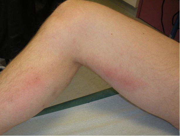
Thrombophlebitis is essentially a composite of two diagnoses: phlebitis, which is a clinical diagnosis in the setting of an erythema and pain overlying a vein and an identified thrombus. In the lower extremities, this is most likely to occur in varicose veins. 

1-Figure1-1.png

In general, risk factors for thrombophlebitis are the same as for DVT (think Virchow's Triad of hypercoagulability, hemodynamic changes (stasis/turbulence), and endothelial injury/dysfunction). These include pregnancy, history of vein excision/ablation, history of prior thrombosis, malignancy, and prior IV catheter placement. 

Special cases:

  • "Mondor": thrombophlebitis of a breast vein, anterior chest vein, or of the dorsal penile vein. The two former should prompt a search for breast cancer and the later is usually due to repetitive trauma

  • "Trousseau's sign of malignancy": migratory thromboembolism, has a strong association with adenocarcinoma of the pancreas and lung


Diagnosis
:
Diagnosis of phlebitis is clinical. Ultrasound should be used to identify a thrombus to distinguish between phlebitis and thrombophlebitis. Patients should also have duplex ultrasound to identify a DVT especially if the area of concern is above the knee. This is important because the rate of concurrent DVT in all cases is 25% and the rate of concurrent PE is 5%. 

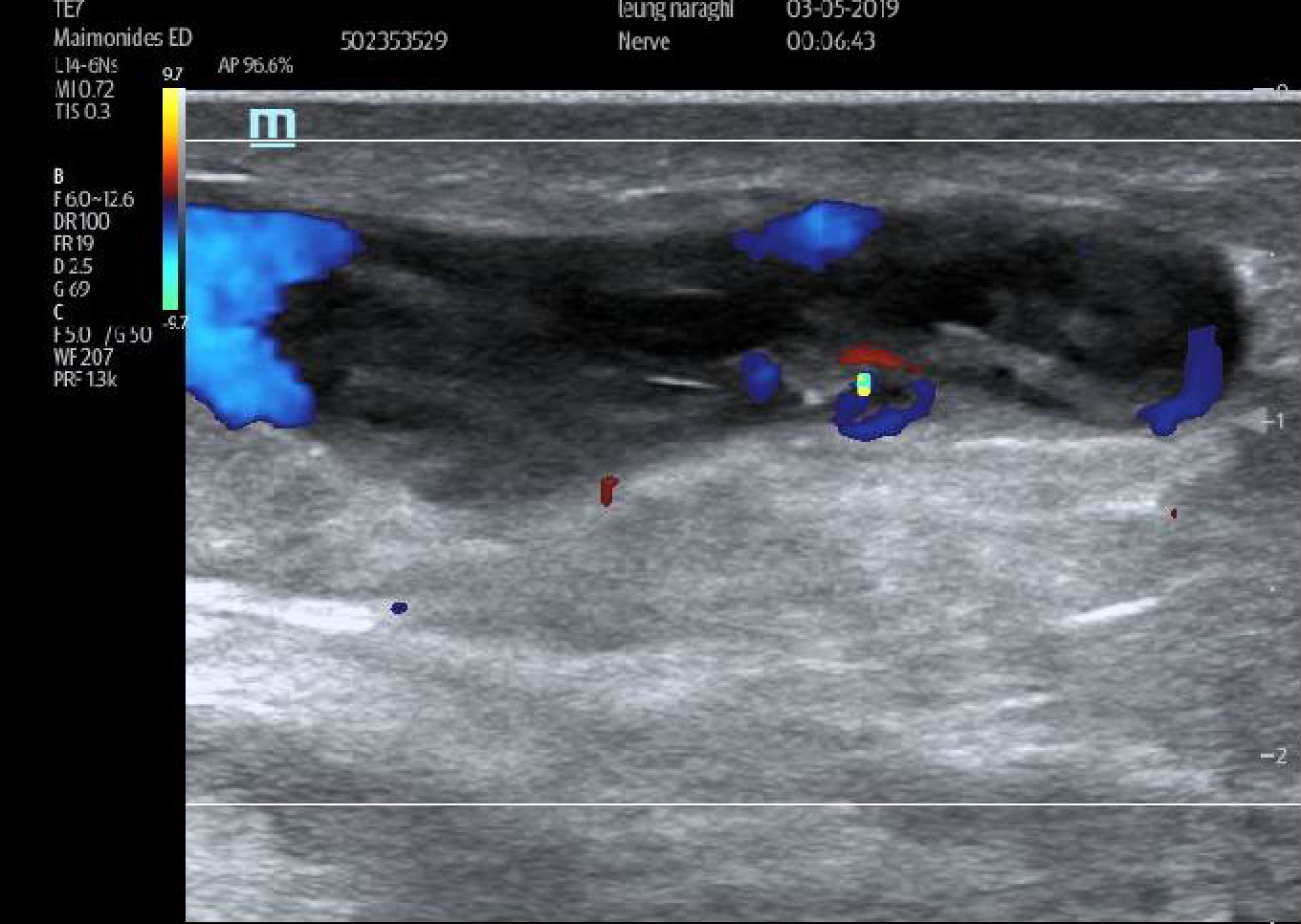
Thrombophlebitis on ultrasound will demonstrate heterogeneous internal echoes within a superficial vein. Unlike an abscess or a lymph node, this will not be discrete and you should be able to trace it out. in the words of the Doge:

"it looks like a weird continuous twisty spaghetti
abscess thing; 
but it's not to be feared, 
mostly superfluous, so get frisky and ultrasound that biddy, 
color doppler, diagnosis, ka-ching". 

There may be flow present (which can help distinguish an abscess from thrombophlebitis; abscess = no flow). Some examples (by the Doge Pologe and myself):

thrombophlebitis.png
Screen Shot 2019-04-15 at 6.57.02 PM.png

Treatment:
As with treatment of below the knee VTE, the treatment of superficial thrombophlebitis is controversial. Patients can be considered low risk (for VTE) if they meet these criteria:

  • Affected vein segment < 5cm

  • Remote from the saphenofemoral/saphenopopliteal junction

  • Low risk for VTE

**Repeat duplex should be obtained in 7-10 days or for worsening symptoms to check for propagation!

NB: thrombophlebitis post-ablation are always low risk and do not require treatment

For these uncomplicated cases, treatment is aimed at alleviating symptoms and prevention of propagation. This includes the following:

  • NSAIDs

  • Warm/cool compresses

  • Elevation of the extremity

  • Compression stockings

Patients that do not qualify as low risk or if they have recurrent thromboembolism should be considered for anticoagulation. Although studies looking at anticoagulation for SVT are small and flawed, NSAIDsLMWH, and fondaparinux have all been shown to decrease incidence of DVT. Patients can also be discharged on Xarelto (this is the only NOAC to be studied for this indication). 

In addition, thromboembolism can become suppurative. Signs and symptoms include high fever (as opposed to the low-grade fever that accompany simple thrombophlebitis) and purulent drainage (duh). In these cases, consider antibiotics


TL;DR

  • Thrombophlebitis = phlebitis (redness/pain along vein) + thrombus

  • Find the thrombus on ultrasound! Look for internal echoes.

  • Low risk patients = below the knee, affected vein < 5cm, distance remove from saphenofemoral/saphenopopliteal junction can be managed with NSAIDs, compression stockings, warm/cool compresses, elevation

  • Low risk patients should get repeat study in 7-10 days to check for propagation

  • High risk patients: consider anticoagulation with LWMH, fondaparinux, or Xarelto

  • Antibiotics for suppurative thrombophlebitis

Special thanks to Dr. Jonas Pologe, Dr. Lawrence Haines, and Dr. Leily Naraghi Bagher Pour. 

Sources:
http://www.emdocs.net/core-em-superficial-venous-thrombosis-svt/
https://www.bmj.com/rapid-response/2011/10/31/superficial-thrombophlebitis-not-straightforward-we-may-think
https://emedicine.medscape.com/article/463256-overview
https://www.uptodate.com/contents/phlebitis-and-thrombosis-of-the-superficial-lower-extremity-veins
https://radiopaedia.org/cases/superficial-thrombophlebitis-1?lang=us

 ·