Failing Up

It as a given that we will fail.  All of us.  Medical or not.  We are all human, which means we make mistakes. So once we accept this as fact, we can then move towards failing better.

When we make a medical error we have a few choices:


1. Live in perpetual doubt

Blame yourself and practice defensive medicine


2. Ignore the error and do nothing, stop caring


3. Failing up – learn from the fail

-       Learning from your medical error takes effort, consideration and time -makes one even more accountable, compassionate and competent.

-       We are all going to fail.

-       When one sweep failures under the carpet, one cannot learn from them effectively and neither can one heal from them effectively

-       Learning to accept this eventuality and incorporating it into our daily life, will allow us to grow as individuals, to more effectively teach others and to take better care of our patients.

-       Strive for post traumatic growth and thriving after failure instead of feelings of shame and isolation.


Failing up strategies:

-       Find a Failure Friend: An empathetic work friend who understands the context; someone who is your safety net and in return you can be their safety net.

-       Be a good Failure Friend to a colleague: Listen/hear them out and empathize/provide affirmation rather than giving advice or solutions. Use reflective listening like you would with a patient and then come up with a joint plan.

-       Teach from your mistakes: Give talks around your difficult cases that incorporate personal strategies on how to cope after the fact, rather than only concentrating on the medical aspects of the case. Talk with peers, residents and medstudents


Watch this excellent FeminEM talk by Dr Sara Gray: https://www.youtube.com/watch?v=aGS6O98p4Q8


References:

https://emergencymedicinecases.com/preventing-burnout-promoting-wellness-emergency-medicine/

https://www.entrepreneur.com/article/331332

https://saragray.org/2017/03/07/failure-friends/

 · 
Share

Pressors in cardiogenic shock

“Pressors” in Cardiogenic Shock in adults


  • Vasopressors- Pure vasoconstriction without any inotropy eg Phenylephrine and Vasopressin

  • Inotrope- Increase cardiac contractility à improving SV and cardiac output without any vasoconstriction eg Milrinone

  • Inopressors - a combination of vasopressors and inotropes, because they lead to both increased cardiac contractility and increased peripheral vasoconstriction eg Norepinephrine, Epinephrine and Dopamine


Norepinephrine- Inopressor

  • Considered the safest Inopressor

  • Less arrhythmogenic than Epinephrine and Dopamine


Mechanism of action

  • Stimulates alpha-1 and alpha-2 receptors

  • Small amount of beta-1 agonist- modest inotropic effect

  • Increased coronary blood flow and afterload

  • Increases venous tone and return with resultant increased preload

Adverse effects

  • Norepinephrine is considered safer than both epinephrine and dopamine.

  • Still carries risk of toxicity to cardiac myocytes, cardiac arrhythmias, and peripheral vasoconstriction leading to tissue ischemia

Indications

  • Norepinephrine is considered first-line in cardiogenic shock with profound hypotension (SBP < 70 mm Hg)

  • Should be used in conjunction with dobutamine in patients with cardiogenic shock and blood pressure higher than 70 mm Hg who fail to respond to dobutamine.

Dosing

  • Use weight-based dosing to avoid the adverse effects associated with norepinephrine use

  • Weight-based dosing is based on GFR

  • Norepinephrine has a rapid onset of action (minutes) and can be titrated every 2-5 minutes


Dobutamine- Inopressor

Mechanism of action

  • Stimulates beta-1 and beta-2 receptors at approximately a 3:1 ratio

  • At high doses (greater than 15 ug/kg/min), dobutamine also becomes a mild alpha-1 agonist.

  • Because it mainly stimulates beta-1 receptors, dobutamine is mostly an inotrope

  • Dobutamine’s stimulation of beta-2 receptors can result in peripheral vasodilation, though the magnitude of this effect is variable à blood pressure in some (but not all) patients.

  • Due to its vasodilatory effects, dobutamine has been shown to improve capillary perfusion independent of changes in blood pressure and cardiac index.

Adverse Effects

  • Studies have demonstrated increased myocardial oxygen demand and malignant arrhythmias typically occuring at doses higher than 15 ug/kg/min

  • Many patients experience hypotension associated with dobutamine use and should be used with caution in patients with systolic blood pressure less than 90 mmHg

  • Dobutamine should only be used in patients with adequate fluid resuscitation


Indications

  • Current ACC/AHA guidelines à first-line agent in management of hypotension associated with acute myocardial infarction

  • But because dobutamine can lower BP, it should only be used if SBP is between 70-100 mmHg, with norepinephrine ready (or already infusing) as well.

  • Dobutamine is typically recommended as the first line agent in cardiogenic shock , but this is not a strong recommendation because several studies have demonstrated benefits to norepinephrine in this setting.

  • If dobutamine is used as a first-line agent, then norepinephrine should be second-line or already infusing, followed by milrinone.


Dosing

  • Dobutamine can be started at 2 mcg/kg/min and titrated to effect, with a maximum dose of 20 mcg/kg/min.

  • Onset of action is 1-2 minutes and the half-life is also approximately 2 minutes à rapidly reversible.


Milrinone- Inodilator

Mechanism of action

  • Milrinone is a phosphodiesterase-3 (PDE3) inhibitor à leads to cardiac smooth muscle relaxation and peripheral vasoconstriction

  • Potent inotropy + diastolic relaxation and vasodilation à to reduced preload, afterload, and systemic vascular resistance (SVR)

  • Milrinone has no beta-adrenergic activity à minimal chronotropic effects.


Adverse Effects

  • Because milrinone decreases preload (and therefore often leads to hypotension), it should only be used in patients who have undergone appropriate fluid resuscitation

  • Use of milrinone often necessitates concurrent vasopressor administration.

  • Because milrinone is metabolized in the kidneys, it should be avoided in patients with renal disease


Indications

  • Recommended for use in patients with daily beta-blocker use and in patients with long-standing heart failure who have developed resistance to catecholamine derivatives

  • Due to PDE’s vasodilatory effect on pulmonary vasculature à theoretical benefit in patients with pulmonary hypertension

Dosing

  • The starting dose of milrinone should ideally be chosen based on that patient’s renal function. The general range is 0.25-0.75 mcg/kg/min.

  • Avoid its use in patients with creatinine clearance less than 50 mL/min.

  • Because of its long onset of action and half-life, milrinone should be titrated every 2 hours (or slower, in the presence of renal disease).


Vasopressin- Pressor

Mechanism of action

  • Vasopressin is an endogenously released hormone (also known as anti-diuretic hormone) à vasopressin receptors in the kidneys à improve GFR

  • Vasopressin receptors on the peripheral vasculature à vasoconstriction.

  • Also causes coronary and cerebral vasodilation


Adverse Effects

  • Vasopressin increases the risk of digital ischemia more significantly than the catecholamine derivatives.

  • No evidence to support the use of vasopressin through a peripheral intravenous line

  • Vasopressin does not have an antidote if extravasation does occur.

Indications

  • Due to its increased risk for digital ischemia à avoid vasopressin in patients with known PVD

  • It has been proposed that because vasopressin leads to coronary vasodilation, it may be a preferable agent in cardiogenic shock but few RCTs investigating vasopressin use in cardiogenic shock.

  • Vasopressin may not lead to pulmonary vasoconstriction à ideal pressor choice in hypotension secondary to pulmonary hypertension à but not enough literature to support routine use in this setting


Dosing


Vasopressin is an endogenous à no utility to titrating vasopressin à used at a set dose of 0.04 U/min, regardless of weight.


Epinephrine- Inopressor


Mechanism of action

  • Beta-1 and beta-2 receptors agonism à more inotropic effects than norepinephrine

  • Epinephrine greatly increases chronotropy (heart rate) and thus stroke volume

  • Some stimulatory effect on alpha-1 receptors

  • Lower doses (1-10 mcg/min) à a beta-1 agonist

  • Higher doses (greater than 10 mcg/min) à an alpha-1 agonist


Adverse effects

  • Associated with an increased risk of tachycardia and lactic acidosis

  • Hyperglycemia

  • Increased incidence of arrhythmogenic events associated with epinephrine

  • More difficult use lactate as a marker of the patient’s response to treatment


Indications

Should be used with extreme caution in cases of cardiogenic shock:

  • RCT of 219 patients with cardiogenic shock found epinephrine to be independently associated with increased 90-day mortality and worsened renal function compared to dobutamine and norepinephrine (not validated).

  • Known increased incidence of arrhythmogenic events associated with epinephrine


Dosing


  • doses of 1-10 mcg/min predominantly activate beta-1 receptors, while doses greater than 10 mcg/min begin to primarily affect alpha-1-mediated vasoconstriction.


Phenylephrine: Not recommended in Cardiogenic shock


Resource: Awesome chart summarizing plessors

 http://www.emdocs.net/wp-content/uploads/2018/02/Inopressor-Summary_chart.pdf


References:

http://www.emdocs.net/evidence-based-approach-pressors-shock-part/

emDOCs.net – Emergency Medicine EducationAn Evidence-Based Approach to Pressors in Shock: Part I - emDOCs.net - Emergency Medicine Education

www.emdocs.net


http://www.emdocs.net/evidence-based-approach-pressors-shock-part-ii/

emDOCs.net – Emergency Medicine EducationAn Evidence-Based Approach to Pressors in Shock: Part II - emDOCs.net - Emergency Medicine Education

www.emdocs.net


Tarvasmäki T, Lassus J, Varpula M, Sionis A, Sund R, Køber L, et al. Current real-life use of vasopressors and inotropes in cardiogenic shock-adrenaline use is associated with excess organ injury and mortality. Critical Care. 2016;20(1):208.

 · 
Share

Trauma in Pregnancy

Resuscitation of the Pregnant Trauma patient


General principles

·      Trauma is the most common cause of non-obstetrical maternal death in the United States

·      Best fetal resuscitation is good maternal resuscitation.

·      Stabilization of the pregnant women is the first priority; then, if the fetus is viable (≥ 23 weeks), fetal heart rate auscultation and fetal monitoring can be initiated and an obstetrical consultation obtained as soon as feasible

·      In Rh-negative pregnant trauma patients, quantification of maternal–fetal hemorrhage by tests such as Kleihauer-Betke should be done to determine the need for additional doses of anti-D immunoglobulin.

·      Tetanus vaccination is safe in pregnancy and should be given when indicated.


Airway

·      Greater risk for difficult intubation than non-pregnant patient

·      Pregnancy related changes à decreased functional residual capacity, reduced respiratory system compliance, increased airway resistance, and increased oxygen requirements

·      Gastric emptying is delayed in pregnancy à greater risk for aspiration

·      Respiratory tract mucosal edema à A smaller size of endotracheal tube is recommended

·      Choice of RSI medications NOT affected by pregnancy status


Breathing

·      Place chest tube one to 2 intercostal spaces higher than usual to account for displacement of the diaphragm during pregnancy

·      Marked increases in basal oxygen consumption à lower threshold for supplemental oxygen


Circulation

·      Fluid and Colloid resuscitation like standard trauma protocol

·      Uteroplacental vasculature is highly responsive to vasopressors, and their administration may decrease placental perfusion à vasopressors should be avoided unless refractory

·      Avoid supine hypotension: Compression of IVC by the uterus can cause up to 30% reduction in cardiac output à Place in left lateral position or by manual displacement of the uterus while the injured patient is secured in the supine position

·      O-negative blood should be transfused in order to avoid Rh sensitization in Rh-negative women

·      Vital signs: heart rate increases by 15% during pregnancy. Tachycardia and hypotension, typical of hypovolemic shock, may appear late in the pregnant trauma patient because of her increased blood volume.

·      Maternal vital signs and perfusion may be preserved at the expense of uteroplacental perfusion, delaying the occurrence of signs of hypovolemic shock

·      Attempt to obtain supra-diaphragmatic intravenous or intraosseous access for volume resuscitation and medication administration.


FAST

·      The FAST is less sensitive for free fluid in the pregnant patient than in non-pregnant patients.  Sensitivity decreases with increasing gestational age, likely due to altered fluid flow within the abdomen.

·      Management of suspected placental abruption should not be delayed pending confirmation by ultrasonography as ultrasound is not a sensitive tool for its diagnosis.


Secondary survey

·      In cases of vaginal bleeding at or after 23 weeks, speculum or digital vaginal examination should be deferred until placenta previa is excluded by a prior or current ultrasound scan.


Imaging

·      Radiographic studies indicated for maternal evaluation including abdominal computed tomography should not be deferred or delayed due to concerns regarding fetal exposure to radiation.

·      Ionizing radiation has the highest teratogenic potential during the period of organogenesis (5–10 weeks), with an increased risk of miscarriage before this period.

·      With abdominal CT during the third trimester the fetal exposure is around 3.5 rads, which is still under the threshold for fetal damage

·      Contrast agents should be used if indicated.


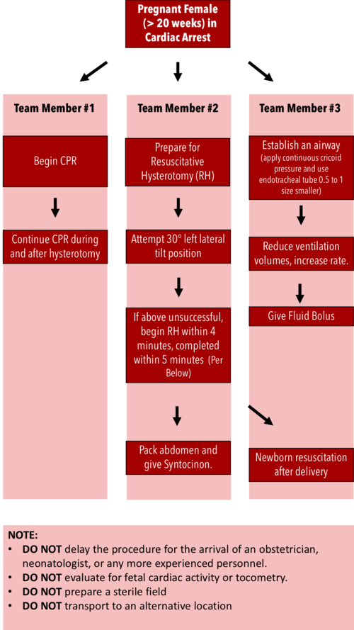
Resuscitative Hysterotomy in Cardiac Arrest

·      Should begin within 4 minutes and completed within 5 minutes of cardiac arrest

·      Both maternal and fetal survival decrease significantly after 5 minutes

·      Do NOT delay the procedure for the arrival of an obstetrician or neonatologist.

·      Do NOT evaluate for fetal cardiac activity or tocometry.

·      Do NOT prepare a sterile field (but be as clean as possible).

·      Do NOT transport to an alternative location.

·      Performing RH increases maternal cardiac output by 30%.


RH Algorithm.png


References:


Tamingthesru.com

EmDocs

Jain, Venu, et al. "Guidelines for the management of a pregnant trauma patient." Journal of Obstetrics and Gynaecology Canada 37.6 (2015): 553-571.

Smith, Kurt A., and Suzanne Bryce. "Trauma in the pregnant patient: an evidence-based approach to management." Emergency medicine practice 15.4 (2013): 1-18.


 · 
Share