Resuscitative TEE

Important: Please complete the Sexual Harassment Online Module ASAP!


Resuscitative Transesophageal echocardiography (TEE)

  • TEE allows the emergency physician to maintain the standard of an ultrasound-informed resuscitation in the scenario of cardiac arrest, where TTE is significantly limited.

  • Focused or resuscitative TEE (4 views) differ from comprehensive TEE (>20 views) that cardiology performs in that it is employed to identify specific questions.

  • TEE allows for potentially shorter chest compression pauses

  • TEE allows for evaluation for the quality of chest compressions

  • TEE allows for visualization of fine V-fib not seen on the monitor


Indications: Cardiac arrest (ACEP)

Contraindications: Esophageal injury or stricture and lack of a definitive airway

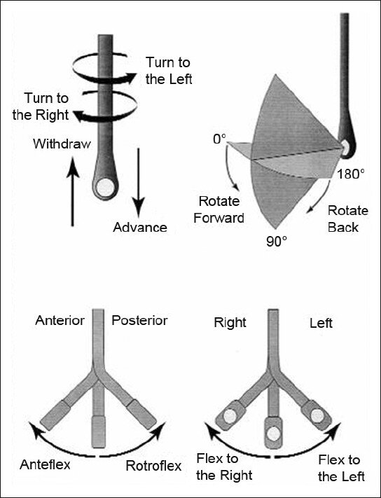
How to manipulate a TEE Probe:

5 different ways you can physically manipulate the TEE probe

1. Withdraw or Advance up or down patient’s esophagus

2. Turn probe to right or left

3. Turn tip of flip in anterior- ante-flexing or in the posterior direction called retro-flexing --> large wheel

4. Turn tip to Left or right -->  small wheel (not typically used for our purposes)

5. In addition, you can rotate the transducer housed within the probe itself (AKA omniplane or multiplane)-->  adjusts the beam angle anywhere between 0° and 180° -->  two smaller buttons ( crystal rotation)

TEE manipulation.jpg


TEE-controls- wheels.png


The views are obtained in the following order: : 

The midesophageal 4-chamber view (ME 4C) is obtained by advancing the TEE probe to the thoracic esophagus and orienting the multiplane at 0-20° in neutral flexion. You may need to retroflex slightly to see all four chambers.

-   The midesophageal long-axis view (ME LAX) is obtained by leaving the probe in the same location as the midesophageal 4-chamber, but increasing the multiplane to between 110° and 160° while in neutral flexion. 

-   The transgastric short axis view (TG- SAX) is obtained by first moving the multiplane to 0°, then advancing the probe into the stomach and ante-flexing the probe

-   The bicaval view (ME bicaval) is obtained by turning the entire probe to the patient’s right towards the superior vena cava (SVC) and inferior vena cava (IVC) while in the mid-esophagus, keeping the multiplane at 90-100° with neutral flexion

 ( The first 3 views are recommended by ACEP. Bicaval not recommended by ACEP) 


TEE views and their analogous TTE views


TTE-and-TEE.gif

Midesophageal four chamber view (ME 4C)

-  Apical four chamber view

-  Great visualization of all chambers as well as the tricuspid and mitral valves in one plane.

-   Evaluation of right and left ventricular systolic function and size

-   Preferred view to evaluate for the presence or absence of a perfusing rhythm during a pulse check.

ME4C.png


Midesophageal Aortic Long Axis view (ME LAX)

-  Midesophageal analogous to the parasternal long axis view in TTE

-   View includes the mitral and aortic valves, as well as the left atrium, left ventricle, and left ventricular outflow tract of the right ventricle.

-   Evaluate left ventricular systolic function, and provides feedback on compression adequacy and location. High-quality compressions cause maximal compression of the left ventricle and visualization of the aortic valve opening and closing indicating forward flow of blood.  Poor quality compressions are seen over the aortic root and there is no valvular indication of forward flow. 

ME- LAX.png


Transgastric Short Axis view (TG- SAX)

- Analogous to the parasternal short axis TTE view

- Evaluate left ventricular systolic function, including any regional wall motion abnormalities

- Can evaluate for acute MI and the presence of septal flattening in this view

TGSAX.png

Mid Esophageal Bicaval View (ME bicaval)

-   Analogous to the inferior vena cava view of TTE

-   Transducer plane cuts through the left atrium (LA), right atrium (RA), IVC and SVC.

This view allows the operator to evaluate for hypovolemia, atrial size, and interatrial septum bowing.

-   Aids in the placement of central venous catheters, transvenous pacemakers, or extracorporeal life support (ECMO) vascular cannulas by observing the initial wire placement in the vasculature

- Can aid inevaluation of fluid status to guide fluid resuscitation (looking at respiratory variation in SVC)

bicaval.png


Pitfalls

-  Compressions do not need to be stopped for TEE insertion. Additionally, the TEE can be left in the esophagus during defibrillation. The probe should be inserted or withdrawn while the tip is in neutral position, and not while the tip is flexed to avoid esophageal injury. 

-  Images should be optimized to avoid foreshortening of the ventricles and to include the appropriate structures for each view.

-  Pericardial effusions must be taken into clinical context, as small effusions can cause tamponade if accumulated rapidly, while large effusions can be well tolerated if they accumulate slowly.

-   Clotted hemopericardium may be isoechoic with the myocardium, making it difficult to identify.

-  Right ventricular failure is not specific to pulmonary embolism, and can be due to pulmonary hypertension or other etiologies such as right sided myocardial infarction, or even cardiac arrest itself.

-  Pleural effusions can be mistaken for pericardial effusions. Multiple views should be used to corroborate findings.

-  Fat pads can be mistaken for pericardial effusions, but these are hypoechoic rather than anechoic and limited to the anterior and apical regions of the heart, not circumferential.


 Resource: 

Check out this 3D module that you can practice on 

https://pie.med.utoronto.ca/TEE/TEE_content/TEE_standardViews_intro.html

References:

Drs Lawrence Haines, Judy Lin and Alyssa Phuoc-Ngyuyen

Images: Adapted from Arntfield R, Pace J, McLeod S, et al. Focused transesophageal echocardiography for emergency physicians-description and results from simulation training of a structured four-view examination. Crit Ultrasound J. 2015;7(1):27.

Teran, Felipe, et al. "Evaluation of out-of-hospital cardiac arrest using transesophageal echocardiography in the emergency department." Resuscitation 137 (2019): 140-147.

EmDocs

ACEP policy statement

https://www.acep.org/patient-care/policy-statements/guidelines-for-the-use-of-transesophageal-echocardiography-tee-in-the-ed-for-cardiac-arrest/

 · 
Share

Non Invasive Ventilation

Non Invasive Ventilation


Definitions:

  • CPAP: applies constant pressure throughout the breathing cycle to increase functional residual capacity (FRC) by recruiting alveoli, decreasing work of breathing, and improving oxygenation.

  • PEEP/EPAP: alveolar pressure before inspiratory flow begins. PEEP à decrease the amount of work required to initiate a breath and decrease atelectasis

  • Bi-level: Cycled ventilation between Inspiratory Positive Airway Pressure (IPAP) and Expiratory Positive Airway Pressure/PEEP. BiPAP supports ventilation and increases oxygenation.

  • Pressure Support: The difference between EPAP and IPAP is referred to as pressure support. Pressure support makes it easier to draw larger tidal volumes


BiPAP/ NIPPV/ Bi-level vs HFNC

·       Oxygenation:  Both devices can almost à  100% FiO2.  HFNC à small PEEP ~5cm, max vs much higher PEEP on NPPV

·       Work of Breathing:  HFNC may wash out the anatomic deadspace à  reduces the work of breathing.  BiPAP can higher pressures and support majority of the work of breathing.

·       Secretion clearance: Important in pneumonia to prevent mucus plugging improve clearance.  BiPAP impairs secretion clearance, whereas HFNC does not seem to.

·       Monitoring: Unable to communicate with patient effectively on BIPAP. BiPAP anxiety provoking and makes it difficult to differentiate between worsening clinical resp status vs anxiety. HFNC facilitates communication  


NIPPV/Bi-level/ BiPAP

COPD

·      Bi-level ventilation à decreases the risk of death (relative risk reduction 48%) and intubation rates (RRR 60%)

·      Number Needed to Treat (NNT) for mortality benefit = 10

·      NNT to prevent intubation = 4

·      Furthermore, when comparing patients with moderate and severe acidosis, bi-level ventilation decreased mortality, rates of intubation, and lengths of stay.

·      ** Ensure patient does not have a PTX that could tension once placed in PPV

Initial Settings:

-       IPAP 8-20 cm H2O (up to 30 cm H20)

-       EPAP 2-6 cm H2O to overcome intrinsic airway collapse

-       Begin with either high IPAP and then titrate down, or low and titrate high.


SCAPE/ CHF exacerbation

·      IPAP assists ventilation à  decreases the WOB

·      EPAP/PEEP increases the FRC by recruiting collapsed alveoli, improving oxygenation, and helping to force interstitial fluid back into the pulmonary vasculature

·      Also, increases intrathoracic pressure à decreased left ventricular (LV) end diastolic volume à decreased afterload and increased LV ejection fraction/stroke volume.

·      Common Initial Settings:

·              IPAP: 10 to 20 cm H20

·              EPAP: 5 to 10 cm H20

·              I:E ratio of IT to ET and is usually set at 1:3 or 1:4 (Inspiratory to Expiratory ratio)

·      Evidence for Bi-level ventilation in CHF exacerbations is unfortunately mostly supportive of CPAP with few trials comparing CPAP and BiPAP

·      Cochrane review looking at NIPPV in CHF exacerbations à CPAP alone has been proven to decrease intubation rates and to decrease in-hospital mortality, without the same benefit seen using bi-level ventilation

·      Lack of evidence does not mean lack of efficacy

BIPAP titration.gif


How to assess your patients on NIV

·       Oxygenation:   good pulse oximetry waveform. ABG is rarely needed to measure oxygenation

·       Work of breathing:  The best metric is the respiratory rate.  Worsening retractions, diaphoresis, tripoding, shallow breathing, and an abdominal paradoxical breathing pattern. 

·       Mentation:  A patient who is easily arousible and mentating adequately doesn't have life-threatening hypercapnia

·       BiPAP screen:  Low tidal volumes and/or low minute ventilation àhypoventilation. But  adequate tidal volumes and minute ventilation suggest a adequate response to NIV

·       Treat the patient, not the ABG- Proven by Brochard 1995 (RCT) investigating the use of BiPAP in COPD:  BiPAP improved mortality despite having no effect on ABG parameters after one hour à  BiPAP can be successful without any immediate effect on the ABG.


High Flow Nasal Cannula

HI-FLOW.png


·       Low flow: NC ~ 1-5 L/min vs NRB ~15 L/min

·       High Flow- 20-60 L/min

·       Normal Resting breathing flow ~15-30 L/min

·       Respiratory distress 60-180 L/min 

  • Adult devices max out at 50-60 L/Min (max it out to start) and the dose for pediatric patient’s (based on trials) is 2L/Kg/Min

  • Maximize your devices flow rate initially then wean the fi02 to maintain your oxygen saturation goal


Adult Indications: Hypoxemic Respiratory failure (mostly Pneumonia) FLORALI Trial, DNR/DNI patients , Pre-oxygenation prior to Intubation


Pediatric Indications: Bronchiolitis, Asthma, Pneumonia, Croup


Take Home Points:

·      Rule out pneumothorax (auscultation, US) prior to placing patients on NIPPV

·      Do not base clinical management solely based off of an ABG esp if patient clinically improving

·      Choose Bi-level vs HFNC based on patient’s diagnosis

·      Best Metric to assess NIV success is respiratory rate


References:


Vital, F. M. R., Ladeira, M. T. & Atallah, A. N. Non-invasive positive pressure ventilation (CPAP or bilevel NPPV) for cardiogenic pulmonary oedema. Cochrane Database Syst. Rev. CD005351 (2013). doi:10.1002/14651858.CD005351.pub3


Frank Lodeserto MD, "High Flow Nasal Cannula (HFNC) – Part 1: How It Works", REBEL EM blog, August 20, 2018. Available at: https://rebelem.com/high-flow-nasal-cannula-hfnc-part-1-how-it-works/.


Frank Lodeserto MD, "High Flow Nasal Cannula (HFNC) – Part 2: Adult & Pediatric Indications", REBEL EM blog, August 23, 2018. Available at: https://rebelem.com/high-flow-nasal-cannula-hfnc-part-2-adult-pediatric-indications/.


EmDocs

EMCrit

 · 
Share

Heparin Anticoagulation in Acute Coronary Syndrome

Heparin Anticoagulation in Acute Coronary Syndrome


STEMI:

·      Anticoagulation for primary PCI addresses 2 pathophysiologic processes: initial thrombin generation caused by spontaneous coronary plaque rupture and secondary thrombin generation caused by iatrogenic introduction of foreign bodies (stents) and arterial dissection (balloon angioplasty)

·      The thrombotic process does not cease on successful implantation of a coronary stent: platelet activation in the setting of endothelial disruption continues and peak ≈2 hours after coronary intervention

·      AHA 2013 Latest Guidelines:

Screen Shot 2019-10-10 at 11.12.57 AM.png


·      European Society of Cardiology guidelines 2017:

Screen Shot 2019-10-10 at 11.12.04 AM.png


·      Both AHA/ ACC and European Society for Cardiology rate LOE as C for UFH but have Class I recommendation

·      Stronger evidence for Bivalirudin and Enoxaparin with either the same Class recommendation or lower

·      No RCTs have been performed since this has been standard of care in the PCI era


Undifferentiated NSTEMI and Unstable Angina:

This is more controversial in terms of choice of agent as well as the therapy but continues to be standard of care with guidelines supporting use of heparin


2014 AHA guidelines for the management of NSTEMI

-       Recommend unfractionated heparin continued for 48 hours or until PCI is performed (LOE B)  

-       With even a higher level of evidence the same guidelines also recommend enoxaparin 1mg/kg subcutaneously every 12 hours with reduced dosing to 1mg/kg subcutaneously in patients with a creatinine clearance <30mL/min) (LOE A)

-       The guidelines recognize that studies supporting this therapy were performed primarily on patients with a diagnosis of unstable angina and in the era before dual anti platelet therapy and early catheterization/revascularization. 

·      Recent retrospective Chinese review published in 2018 concluded that parenteral anticoagulation therapy did not decrease mortality in patients with NSTEMI undergoing PCI but did have more bleeding events compared to non-parenteral anticoagulation therapy

·      Cochrane review à patients treated with heparins had a similar risk of mortality, revascularization and recurrent angina. However, those treated with heparins had a decreased risk of myocardial infarction (driven by the largest study (FRISC), and they used the 6 day outcome of that trial, rather than the 40 or 150 day outcomes that we know were negative) this was based on a higher incidence of minor bleeding. 

·      Shared decision making can be employed in this setting

·      The risk versus benefit profile might be different for patient who have a history of GIB, known brain aneurysm, high fall risk patients etc


NSTEMI with noninvasive management


·      Rebound effect- Heparin causes a transient reduction in MI rates, with a rebound in infarction after anticoagulation is withdrawn.   There is no long-term sustained mortality benefit in literature

·      Anticoagulation with heparin exposes the patient to a risk of hemorrhage without any known long-term benefit evidenced in literature.  

·      Furthermore, heparin delays the occurrence of ischemic events to a later time-point in their hospital course, when the patient may be less closely monitored.

·      The potential benefit heparin “bridging” to definitive therapy is not realized when patients’ are managed medically and don’t receive an intervention (PCI or CABG)

·      Decision to anticoagulated can be deferred to the consultants managing the patient as they would be more aware of the likely management of the patient with an intervention.


Special considerations:


Instent thrombosis- Benefit anticoagulation as secondary thrombin generation caused by iatrogenic introduction of foreign bodies

Effect of Hemodialysis on troponin levels in ESRD patients - “Troponinemia” could reflect chronic microinfarctions or correlate with left ventricular hypertrophy. HD process itself might cause undesirable myocardial injury and enhance post HD TnI levels. The effect of HD on TnI levels are unestablished, reporting either increasing, unchanged or decreasing of post-dialysis hsTnI levels 

Rhabdomyolysis- The prevalence of false positive cTnI in the ED patients with rhabdomylolyiss was 17% in one study although there is significant paucity of evidence.


Reference:

REBELEM

PulmCrit

First10EM


Amsterdam, Ezra A., et al. "2014 AHA/ACC guideline for the management of patients with non–ST-elevation acute coronary syndromes: a report of the American College of Cardiology/American Heart Association Task Force on Practice Guidelines." Journal of the American College of Cardiology 64.24 (2014): e139-e228.


Ibanez, Borja, et al. "2017 ESC Guidelines for the management of acute myocardial infarction in patients presenting with ST-segment elevation: The Task Force for the management of acute myocardial infarction in patients presenting with ST-segment elevation of the European Society of Cardiology (ESC)." European heart journal 39.2 (2017): 119-177.


Dauerman, Harold L. "Anticoagulation Strategies for Primary Percutaneous Coronary Intervention." (2015).


Li, Siu Fai, Jennifer Zapata, and Elizabeth Tillem. "The prevalence of false-positive cardiac troponin I in ED patients with rhabdomyolysis." The American journal of emergency medicine 23.7 (2005): 860-863.


Tarapan, Tanawat, et al. "High sensitivity Troponin-I levels in asymptomatic hemodialysis patients." Renal failure 41.1 (2019): 393-400.


Chen, JY et al. Association of Parenteral Anticoagulation Therapy With Outcomes in Chinese Patients Undergoing Percutaneous Coronary Intervention for Non-ST-Segment Elevation Acute Coronary Syndrome. JAMA Intern Med 2018. PMID: 30592483


 · 
Share