POTD: Revisiting the Resuscitative Hysterotomy

Formerly known as the perimortem cesarean section, the resuscitative hysterotomy is performed in a pregnant patient of > 20 weeks gestation in cardiac arrest to improve the chances of ROSC.


Forget about the 4-Minute and 5-Minute rule!

  • Even in controlled simulations with obstetric teams, timing has been problematic.

  • While the procedure should be performed as quickly as possible to improve outcomes, there is generally no contraindication to performing the procedure beyond the 5 minute mark.

  • The procedure has benefited pregnant patients up to 15 minutes and fetuses up to 30 minutes after maternal cardiac arrest.


DO’s and DONT’s

  • DO assign team roles and prepare all equipment prior to patient arrival

  • DO start chest compressions immediately, establish an airway, and get IV access

  • DO give fluids

  • DO give blood in the setting of trauma

  • DO NOT stop to evaluate for fetal cardiac activity or tocometry

  • DO NOT prepare a sterile field (but be as clean as possible)

  • DO NOT wait for OB/GYN to arrive before starting the procedure

  • DO NOT transport the patient to another location


THE PROCEDURE

  1. Drench the abdomen in betadine and get ready to cut

  2. With a scalpel, make a vertical incision from the xiphoid process down to the pubic symphysis, cutting through the skin, fat, fascia, and peritoneum

  3. Avoid cutting the bladder — find it, and retract it

  4. Blunt dissect down to the uterus

  5. Make a vertical incision in the uterus large enough to fit 2 fingers in

  6. Once inside, lift the uterine wall with your fingers

  7. Use blunt scissors to divide the uterus between your fingers and extend the incision

  8. Deliver the fetus

  9. Double clamp the umbilical cord and cut BETWEEN the clamps

  10. Deliver the placenta

  11. Wipe the endometrial cavity clean with a clean, moist lap pad

  12. Pack the uterine cavity with sterile towels

  13. Continue resuscitation

WATCH EMCrit VIDEO of a LIVE SIMULATED RESUSCITATIVE HYSTEROTOMY
https://www.youtube.com/watch?v=IwDWv2iyAos

SOURCES

  1. Tintinalli’s Emergency Medicine, 9th Edition pp 646-647

  2. Rose, C.H. et al, Challenging the 4- to 5-minute Rule: From Perimortem Cesarean to Resuscitative Hysterotomy Obstetric Anesthesia Digest December 2016 - Volume 36 - Issue 4 - p 171

  3. WikiEM: Resuscitative Hysterotomy


 · 

Trauma in Pregnancy

Resuscitation of the Pregnant Trauma patient


General principles

·      Trauma is the most common cause of non-obstetrical maternal death in the United States

·      Best fetal resuscitation is good maternal resuscitation.

·      Stabilization of the pregnant women is the first priority; then, if the fetus is viable (≥ 23 weeks), fetal heart rate auscultation and fetal monitoring can be initiated and an obstetrical consultation obtained as soon as feasible

·      In Rh-negative pregnant trauma patients, quantification of maternal–fetal hemorrhage by tests such as Kleihauer-Betke should be done to determine the need for additional doses of anti-D immunoglobulin.

·      Tetanus vaccination is safe in pregnancy and should be given when indicated.


Airway

·      Greater risk for difficult intubation than non-pregnant patient

·      Pregnancy related changes à decreased functional residual capacity, reduced respiratory system compliance, increased airway resistance, and increased oxygen requirements

·      Gastric emptying is delayed in pregnancy à greater risk for aspiration

·      Respiratory tract mucosal edema à A smaller size of endotracheal tube is recommended

·      Choice of RSI medications NOT affected by pregnancy status


Breathing

·      Place chest tube one to 2 intercostal spaces higher than usual to account for displacement of the diaphragm during pregnancy

·      Marked increases in basal oxygen consumption à lower threshold for supplemental oxygen


Circulation

·      Fluid and Colloid resuscitation like standard trauma protocol

·      Uteroplacental vasculature is highly responsive to vasopressors, and their administration may decrease placental perfusion à vasopressors should be avoided unless refractory

·      Avoid supine hypotension: Compression of IVC by the uterus can cause up to 30% reduction in cardiac output à Place in left lateral position or by manual displacement of the uterus while the injured patient is secured in the supine position

·      O-negative blood should be transfused in order to avoid Rh sensitization in Rh-negative women

·      Vital signs: heart rate increases by 15% during pregnancy. Tachycardia and hypotension, typical of hypovolemic shock, may appear late in the pregnant trauma patient because of her increased blood volume.

·      Maternal vital signs and perfusion may be preserved at the expense of uteroplacental perfusion, delaying the occurrence of signs of hypovolemic shock

·      Attempt to obtain supra-diaphragmatic intravenous or intraosseous access for volume resuscitation and medication administration.


FAST

·      The FAST is less sensitive for free fluid in the pregnant patient than in non-pregnant patients.  Sensitivity decreases with increasing gestational age, likely due to altered fluid flow within the abdomen.

·      Management of suspected placental abruption should not be delayed pending confirmation by ultrasonography as ultrasound is not a sensitive tool for its diagnosis.


Secondary survey

·      In cases of vaginal bleeding at or after 23 weeks, speculum or digital vaginal examination should be deferred until placenta previa is excluded by a prior or current ultrasound scan.


Imaging

·      Radiographic studies indicated for maternal evaluation including abdominal computed tomography should not be deferred or delayed due to concerns regarding fetal exposure to radiation.

·      Ionizing radiation has the highest teratogenic potential during the period of organogenesis (5–10 weeks), with an increased risk of miscarriage before this period.

·      With abdominal CT during the third trimester the fetal exposure is around 3.5 rads, which is still under the threshold for fetal damage

·      Contrast agents should be used if indicated.


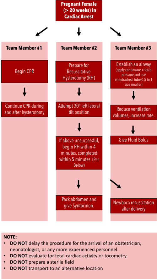
Resuscitative Hysterotomy in Cardiac Arrest

·      Should begin within 4 minutes and completed within 5 minutes of cardiac arrest

·      Both maternal and fetal survival decrease significantly after 5 minutes

·      Do NOT delay the procedure for the arrival of an obstetrician or neonatologist.

·      Do NOT evaluate for fetal cardiac activity or tocometry.

·      Do NOT prepare a sterile field (but be as clean as possible).

·      Do NOT transport to an alternative location.

·      Performing RH increases maternal cardiac output by 30%.


RH Algorithm.png


References:


Tamingthesru.com

EmDocs

Jain, Venu, et al. "Guidelines for the management of a pregnant trauma patient." Journal of Obstetrics and Gynaecology Canada 37.6 (2015): 553-571.

Smith, Kurt A., and Suzanne Bryce. "Trauma in the pregnant patient: an evidence-based approach to management." Emergency medicine practice 15.4 (2013): 1-18.


 · 

Case of eclampsia in your resus bay

Diagnosis:

  • new-onset tonic-clonic, focal, or multifocal seizures in the absence of other causative conditions (eg, epilepsy, cerebral arterial ischemia and infarction, intracranial hemorrhage, drug use), 

  • typically but does not have to be present in the presence of preexisting hypertensive disorder of pregnancy (preeclampsia, gestational hypertension, HELLP syndrome)


Presentation:

  • Hypertension 

  • Headache (persistent frontal or occipital headaches or thunderclap headaches)

  • Visual disturbances (scotomata, loss of vision [cortical blindness], blurred vision, diplopia, visual field defects [eg, homonymous hemianopsia], photophobia)

  • Right upper quadrant or epigastric pain 

  • Asymptomatic 


Management:

  • Start with ABCs

  • Consider alternative causes of seizures based on additional information other than eclampsia: hyponatremia, ICH, hypoglycemia, etc.

  • Usually eclamptic seizures subside on its own

  • If pt is seizing => administer Mg Loading dose 4-6 g IV over 15 to 20 minutes. An alternative dose/route is magnesium sulfate 5 g intramuscularly into each buttock for a total of 10 g

  • Followed by Maintenance dose – magnesium sulfate 2 g/hour as a continuous IV infusion to women with good renal function.

  • If pt is in status => in cases refractory to magnesium sulfate (patient is still seizing at 20 minutes after the bolus or more than two recurrences), administer sodium amobarbital (250 mg IV over three minutes), thiopental, or phenytoin (1250 mg IV at a rate of 50 mg/minute). In this case pt will need to be intubated.

If need to consider intubation:

  • Medications:

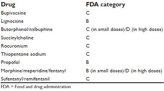
    • Induction - consider propofol (category B)

      • You want to avoid: Etomidate - lowers seizure threshold and Ketamine - worsens HTN


    • Paralytics - rocuronium or succinylcholine, yet both of the medications are category C so use minimal dose to reach the desired effect, avoid additional doses

pregnancy medications.jpg


Next consider hypertensive control if BP diastolic pressures greater than 105 to 110 mmHg or systolic blood pressures ≥160 mmHg:

  • Labetalol - 20 mg IV gradually over 2 minutes.

  • Hydralazine - 5 mg IV gradually over 1 to 2 minutes.

  • Nifedipine immediate release - 10 mg orally.

  • Nicardipine (parenteral) - The initial dose is 5 mg/hour intravenously by infusion pump and can be increased to a maximum of 15 mg/hour.

Proceed with labs, consider HELLP syndrome labs, type and screen, fluids. 

Call OB/GYN early

The definitive treatment for eclampsia is prompt delivery.