Trauma in Pregnancy

Resuscitation of the Pregnant Trauma patient


General principles

·      Trauma is the most common cause of non-obstetrical maternal death in the United States

·      Best fetal resuscitation is good maternal resuscitation.

·      Stabilization of the pregnant women is the first priority; then, if the fetus is viable (≥ 23 weeks), fetal heart rate auscultation and fetal monitoring can be initiated and an obstetrical consultation obtained as soon as feasible

·      In Rh-negative pregnant trauma patients, quantification of maternal–fetal hemorrhage by tests such as Kleihauer-Betke should be done to determine the need for additional doses of anti-D immunoglobulin.

·      Tetanus vaccination is safe in pregnancy and should be given when indicated.


Airway

·      Greater risk for difficult intubation than non-pregnant patient

·      Pregnancy related changes à decreased functional residual capacity, reduced respiratory system compliance, increased airway resistance, and increased oxygen requirements

·      Gastric emptying is delayed in pregnancy à greater risk for aspiration

·      Respiratory tract mucosal edema à A smaller size of endotracheal tube is recommended

·      Choice of RSI medications NOT affected by pregnancy status


Breathing

·      Place chest tube one to 2 intercostal spaces higher than usual to account for displacement of the diaphragm during pregnancy

·      Marked increases in basal oxygen consumption à lower threshold for supplemental oxygen


Circulation

·      Fluid and Colloid resuscitation like standard trauma protocol

·      Uteroplacental vasculature is highly responsive to vasopressors, and their administration may decrease placental perfusion à vasopressors should be avoided unless refractory

·      Avoid supine hypotension: Compression of IVC by the uterus can cause up to 30% reduction in cardiac output à Place in left lateral position or by manual displacement of the uterus while the injured patient is secured in the supine position

·      O-negative blood should be transfused in order to avoid Rh sensitization in Rh-negative women

·      Vital signs: heart rate increases by 15% during pregnancy. Tachycardia and hypotension, typical of hypovolemic shock, may appear late in the pregnant trauma patient because of her increased blood volume.

·      Maternal vital signs and perfusion may be preserved at the expense of uteroplacental perfusion, delaying the occurrence of signs of hypovolemic shock

·      Attempt to obtain supra-diaphragmatic intravenous or intraosseous access for volume resuscitation and medication administration.


FAST

·      The FAST is less sensitive for free fluid in the pregnant patient than in non-pregnant patients.  Sensitivity decreases with increasing gestational age, likely due to altered fluid flow within the abdomen.

·      Management of suspected placental abruption should not be delayed pending confirmation by ultrasonography as ultrasound is not a sensitive tool for its diagnosis.


Secondary survey

·      In cases of vaginal bleeding at or after 23 weeks, speculum or digital vaginal examination should be deferred until placenta previa is excluded by a prior or current ultrasound scan.


Imaging

·      Radiographic studies indicated for maternal evaluation including abdominal computed tomography should not be deferred or delayed due to concerns regarding fetal exposure to radiation.

·      Ionizing radiation has the highest teratogenic potential during the period of organogenesis (5–10 weeks), with an increased risk of miscarriage before this period.

·      With abdominal CT during the third trimester the fetal exposure is around 3.5 rads, which is still under the threshold for fetal damage

·      Contrast agents should be used if indicated.


Resuscitative Hysterotomy in Cardiac Arrest

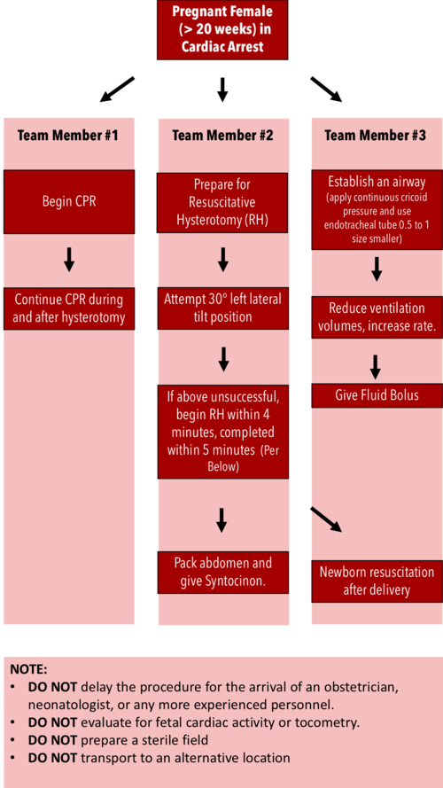
·      Should begin within 4 minutes and completed within 5 minutes of cardiac arrest

·      Both maternal and fetal survival decrease significantly after 5 minutes

·      Do NOT delay the procedure for the arrival of an obstetrician or neonatologist.

·      Do NOT evaluate for fetal cardiac activity or tocometry.

·      Do NOT prepare a sterile field (but be as clean as possible).

·      Do NOT transport to an alternative location.

·      Performing RH increases maternal cardiac output by 30%.


RH Algorithm.png


References:


Tamingthesru.com

EmDocs

Jain, Venu, et al. "Guidelines for the management of a pregnant trauma patient." Journal of Obstetrics and Gynaecology Canada 37.6 (2015): 553-571.

Smith, Kurt A., and Suzanne Bryce. "Trauma in the pregnant patient: an evidence-based approach to management." Emergency medicine practice 15.4 (2013): 1-18.


 · 

Resuscitative TEE

Important: Please complete the Sexual Harassment Online Module ASAP!


Resuscitative Transesophageal echocardiography (TEE)

  • TEE allows the emergency physician to maintain the standard of an ultrasound-informed resuscitation in the scenario of cardiac arrest, where TTE is significantly limited.

  • Focused or resuscitative TEE (4 views) differ from comprehensive TEE (>20 views) that cardiology performs in that it is employed to identify specific questions.

  • TEE allows for potentially shorter chest compression pauses

  • TEE allows for evaluation for the quality of chest compressions

  • TEE allows for visualization of fine V-fib not seen on the monitor


Indications: Cardiac arrest (ACEP)

Contraindications: Esophageal injury or stricture and lack of a definitive airway

How to manipulate a TEE Probe:

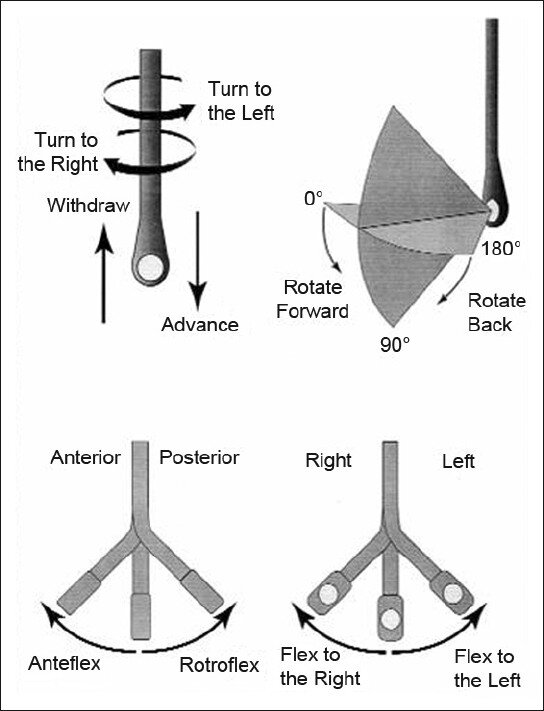
5 different ways you can physically manipulate the TEE probe

1. Withdraw or Advance up or down patient’s esophagus

2. Turn probe to right or left

3. Turn tip of flip in anterior- ante-flexing or in the posterior direction called retro-flexing --> large wheel

4. Turn tip to Left or right -->  small wheel (not typically used for our purposes)

5. In addition, you can rotate the transducer housed within the probe itself (AKA omniplane or multiplane)-->  adjusts the beam angle anywhere between 0° and 180° -->  two smaller buttons ( crystal rotation)

TEE manipulation.jpg


TEE-controls- wheels.png


The views are obtained in the following order: : 

The midesophageal 4-chamber view (ME 4C) is obtained by advancing the TEE probe to the thoracic esophagus and orienting the multiplane at 0-20° in neutral flexion. You may need to retroflex slightly to see all four chambers.

-   The midesophageal long-axis view (ME LAX) is obtained by leaving the probe in the same location as the midesophageal 4-chamber, but increasing the multiplane to between 110° and 160° while in neutral flexion. 

-   The transgastric short axis view (TG- SAX) is obtained by first moving the multiplane to 0°, then advancing the probe into the stomach and ante-flexing the probe

-   The bicaval view (ME bicaval) is obtained by turning the entire probe to the patient’s right towards the superior vena cava (SVC) and inferior vena cava (IVC) while in the mid-esophagus, keeping the multiplane at 90-100° with neutral flexion

 ( The first 3 views are recommended by ACEP. Bicaval not recommended by ACEP) 


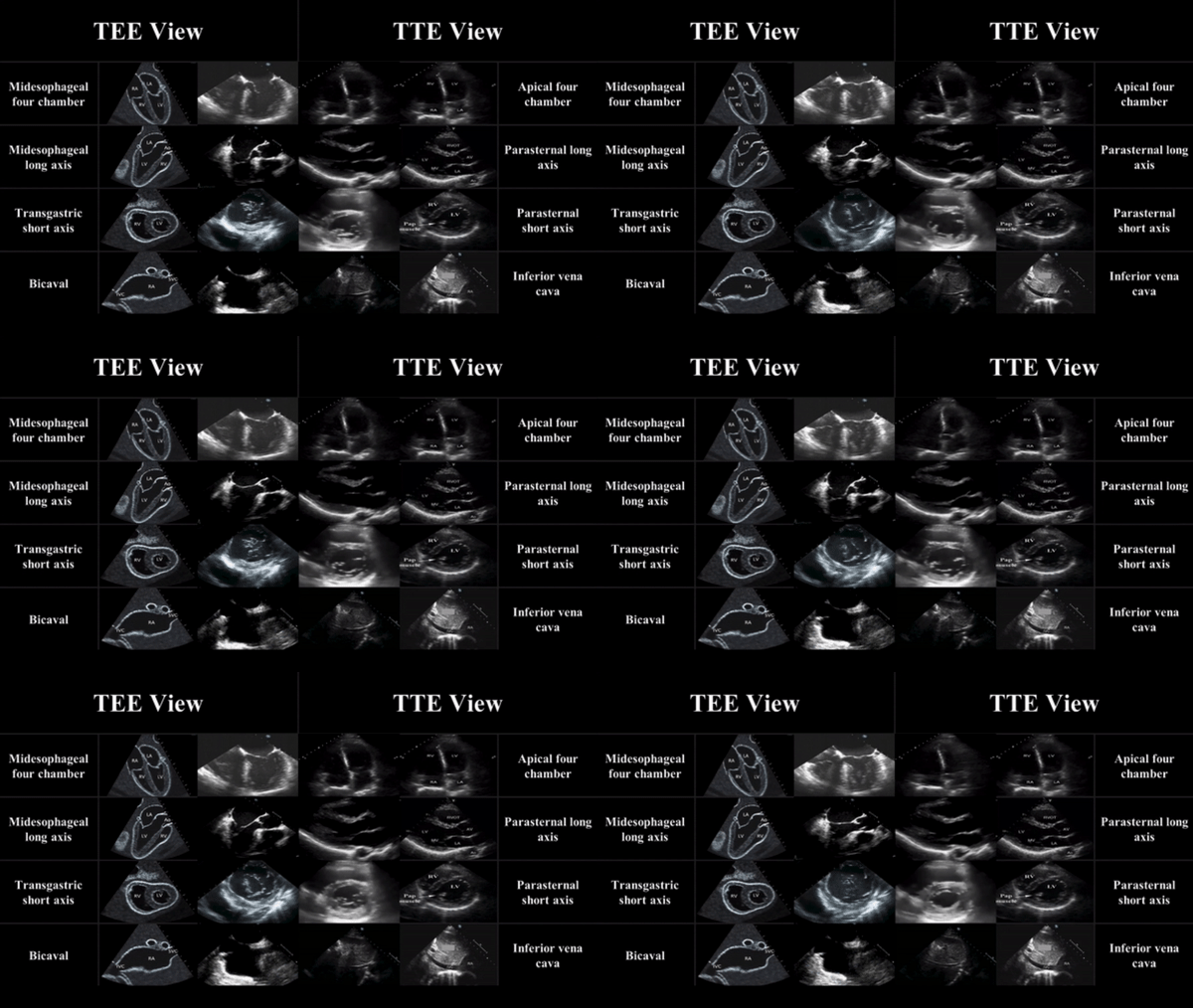
TEE views and their analogous TTE views


TTE-and-TEE.gif

Midesophageal four chamber view (ME 4C)

-  Apical four chamber view

-  Great visualization of all chambers as well as the tricuspid and mitral valves in one plane.

-   Evaluation of right and left ventricular systolic function and size

-   Preferred view to evaluate for the presence or absence of a perfusing rhythm during a pulse check.

ME4C.png


Midesophageal Aortic Long Axis view (ME LAX)

-  Midesophageal analogous to the parasternal long axis view in TTE

-   View includes the mitral and aortic valves, as well as the left atrium, left ventricle, and left ventricular outflow tract of the right ventricle.

-   Evaluate left ventricular systolic function, and provides feedback on compression adequacy and location. High-quality compressions cause maximal compression of the left ventricle and visualization of the aortic valve opening and closing indicating forward flow of blood.  Poor quality compressions are seen over the aortic root and there is no valvular indication of forward flow. 

ME- LAX.png


Transgastric Short Axis view (TG- SAX)

- Analogous to the parasternal short axis TTE view

- Evaluate left ventricular systolic function, including any regional wall motion abnormalities

- Can evaluate for acute MI and the presence of septal flattening in this view

TGSAX.png

Mid Esophageal Bicaval View (ME bicaval)

-   Analogous to the inferior vena cava view of TTE

-   Transducer plane cuts through the left atrium (LA), right atrium (RA), IVC and SVC.

This view allows the operator to evaluate for hypovolemia, atrial size, and interatrial septum bowing.

-   Aids in the placement of central venous catheters, transvenous pacemakers, or extracorporeal life support (ECMO) vascular cannulas by observing the initial wire placement in the vasculature

- Can aid inevaluation of fluid status to guide fluid resuscitation (looking at respiratory variation in SVC)

bicaval.png


Pitfalls

-  Compressions do not need to be stopped for TEE insertion. Additionally, the TEE can be left in the esophagus during defibrillation. The probe should be inserted or withdrawn while the tip is in neutral position, and not while the tip is flexed to avoid esophageal injury. 

-  Images should be optimized to avoid foreshortening of the ventricles and to include the appropriate structures for each view.

-  Pericardial effusions must be taken into clinical context, as small effusions can cause tamponade if accumulated rapidly, while large effusions can be well tolerated if they accumulate slowly.

-   Clotted hemopericardium may be isoechoic with the myocardium, making it difficult to identify.

-  Right ventricular failure is not specific to pulmonary embolism, and can be due to pulmonary hypertension or other etiologies such as right sided myocardial infarction, or even cardiac arrest itself.

-  Pleural effusions can be mistaken for pericardial effusions. Multiple views should be used to corroborate findings.

-  Fat pads can be mistaken for pericardial effusions, but these are hypoechoic rather than anechoic and limited to the anterior and apical regions of the heart, not circumferential.


 Resource: 

Check out this 3D module that you can practice on 

https://pie.med.utoronto.ca/TEE/TEE_content/TEE_standardViews_intro.html

References:

Drs Lawrence Haines, Judy Lin and Alyssa Phuoc-Ngyuyen

Images: Adapted from Arntfield R, Pace J, McLeod S, et al. Focused transesophageal echocardiography for emergency physicians-description and results from simulation training of a structured four-view examination. Crit Ultrasound J. 2015;7(1):27.

Teran, Felipe, et al. "Evaluation of out-of-hospital cardiac arrest using transesophageal echocardiography in the emergency department." Resuscitation 137 (2019): 140-147.

EmDocs

ACEP policy statement

https://www.acep.org/patient-care/policy-statements/guidelines-for-the-use-of-transesophageal-echocardiography-tee-in-the-ed-for-cardiac-arrest/

 · 

Indications for use of Tranexamic Acid (TXA)

Indications for use of Tranexamic Acid (TXA)

Trauma


Trial Name: CRASH 2 (Positive trial)

Trial Type: Multicenter, double-blind RCT

Sample size: 20,211

Dose/Route of TXA: Loading dose 1g over 10 min, then infusion of 1g over 8hr

Primary outcome: All-cause mortality within 4 weeks of injury

Secondary outcome: Vascular occlusive events (AMI, stroke, PE, and DVT), surgical intervention, receipt of blood transfusion, and units of blood products transfused

Results:  Reduced All-cause mortality p 0.0035, death due to hemorrhage p 0.0077, no significant vascular occlusion p 0.96

Risk of thrombotic events: No increase in risk

Take home point: The use of TXA in trauma patients with “significant bleeding” reduces all-cause mortality without an increase in thromboembolic events.  This effect seems to be greatest in the subset of patients with severe shock (SBP ≤70mmHg) and when given ≤3 hours from time of injury 


Shakur H et al. Effects of Tranexamic Acid on Death, Vascular Occlusive Events, and Blood Transfusion in Trauma Patients with Significant Haemorrhage. Lancet 2010. PMID: 20554319


Trial Name: MATTERs (Positive trial)

Trial Type: Single center, retrospective, observational study

Sample size: 896

Dose/ route of TXA: 1 g initially, 2nd dose per MD discretion

Primary outcome: 24hr mortality, 48hr mortality, and 30-day mortality

Secondary outcome: Transfusion requirements and rate of thromboembolic complications.

Results: Not significantly decreased 24 hr p >0.05, Significantly decreased 48hrs p 0.004 and 30 day mortality p 0.03

Risk of thrombotic events: Increased overall VTE p 0.001 but patients who had a VTE also had higher burden of injury

Take home point:  Patients with penetrating injuries, requiring blood transfusions within 1hr of presentation the use of TXA reduced overall mortality


Morrison JJ et al. Military Application of Tranexamic Acid in Trauma Emergency Resuscitation (MATTERs) Study. Arch Surg 2012. PMID: 22006852


ICH


Trial Name: Meta-Analysis of TXA for Traumatic Brain Injury- negative trial

Trial Type: Meta-analysis and systematic review of RCTs or quasi-RCTs 

Sample size:  510

Outcome: Mortality, neurological function, hematoma expansion

Results: statistically significant reduction in ICH progression with TXA non-statistically significant improvement of clinical outcomes in ED patients with TBI.

Risk of thrombotic events: No adverse effects reported

Take home point: Did not lead to a statistically significant mortality benefit or improved neurological functional status. Further evidence is required to support its routine use in patients with TBI.


Zehtabchi S et al. Tranexamic Acid for Traumatic Brain Injury: A Systematic Review and Meta-Analysis. Am J Emerg Med 2014. PMID: 25447601


Trial Name: Tranexamic Acid for Hyperacute Primary IntraCerebral Haemorrhage (TICH-2)- Negative

Trial Type: International, randomized, double-blind, placebo-controlled, parallel group

Sample size:  2325

Dose of TXA used: 1g IV TXA bolus followed by an 8hr infusion of 1g of TXA 

Outcome:  Functional Status at Day 90, Hematoma Expansion at Day 2, Mean Hematoma Volume Expansion from Baseline to 24hr, Death by Day 7, Death by Day 90

Results: No difference in neurological impairment (mean NIHSS score at day 7), 90-day functional outcomes, length of hospital stay, discharge disposition, venous thromboembolic events, or arterial occlusions

Risk of thrombotic events: None

Take home point: TXA was given >3hrs after stroke onset, patients had more severe strokes, and larger hematoma volumes (>60mLs) than prior studies. Possible benefit if given to a subset of patient within 3 hours with smaller strokes but cannot be recommended at this time in clinical practice for spontaneous ICH based on the results of these trials


Sprigg N et al. Tranexamic Acid for Hyperacute Primary IntraCerebral Haemorrhage (TICH-2): An International Randomised, Placebo-Controlled, Phase 3 Superiority Trial. Lancet 2018. PMID: 29778325


Post Partum Hemorrhage


Trial Name: WOMAN trial – Negative trial

Trial Type: Randomized, double-blind, placebo-controlled trial,

Sample size:  20,060 ≥16 years of age with post-partum hemorrhage after vaginal delivery or caesarean section 

Dose of TXA used: 1 g IV vs matching placebo, If bleeding continued after 30 minutes or stopped and restarted within 24hrs, a second dose of 1g of TXA or placebo was given

Outcome: Initial outcome of all-cause mortality and/or hysterectomy within 42 days of giving birth

Final Primary Outcome: Death from PPH

Results: No difference in all cause mortality or hysterctomy

Risk of thrombotic events:

Take home point: It is difficult to draw definitive conclusions from this trial as the NNT was still large (i.e. ≈250) and the study had a fragility index of 0. Data showed a consistent association of delayed administration of TXA with no benefit

WOMAN Trial Collaborators. Effect of Early Tranexamic Acid Administration on Mortality, Hysterectomy, and Other Morbidities in Women with Post-Partum Haemorrhage (WOMAN): An International, Randomised, Double-Blind, Placebo-Controlled Trial. Lancet 2017. PMID: 28456509


UGIB


Trial Name: Cochrane review

Trial Type: Systematic review and meta-analysis of 8 RCTs

Sample size:  1700

Dose of TXA used: Total daily dose of TXA ranged from 4 – 8g and ranged from 2 – 7 days with both PO and IV adminsteration

Outcome: Primary: all-cause mortality and adverse events

Secondary: Rebleeding and surgery

Results: All-Cause Mortality p 0.007, rebleeding P = 0.07

Risk of thrombotic events: No difference in thromboembolic events (only evaluated in 4 trials)

Take home point: May benefit in higher risk patients but better RCTs required to confirm or refute evidence. HALT IT trial underway currently with N of 12000


Bennett C et al. Tranexamic Acid for Upper Gastrointestinal Bleeding (Review). Cochrane Database Syst Rev 2014. PMID: 25414987


Epistaxis


Trial Name: Zahed et al 2017 – Positive study

Trial Type: Randomized, parallel group clinical trial

Sample size:  124 on antiplatelets

Dose of TXA used: topical TXA (500mg in 5mL) or anterior nasal packing.

Outcome: Primary outcome resolution at 10 minutes. Secondary outcomes were re-bleeding rate at 24hours and one week, ED length of stay, and patient satisfaction

Results: epistaxis treatment with topical application of TXA resulted in faster bleeding cessation (NNT 2) , less re-bleeding at 1-week, shorter ED LOS, and higher patient satisfaction as compared with anterior nasal packing.

Risk of thrombotic events: not evaluated

Take home point: Do it!

Zahed R et al. Topical Tranexamic Acid Compared With Anterior Nasal Packing or Treatment of Epistaxis in Patients Taking Antiplatelet Drugs: Randomized Controlled Trial. Acad Emerg Med 2017. PMID: 29125679


Post-Tonsillectomy Bleeding


Trial Name: Meta-Analysis 2012

Trial Type: Systematic review and meta-analysis

Sample size:  7 studies with 2,444 patients

Dose of TXA used: 250mg for children <25kg, 500mg for children >25kg

Outcome: mean volume of blood loss

Results: TXA led to a significant reduction of tonsillectomy blood loss volume but had no impact on the rate of patients with post-tonsillectomy hemorrhage.

Risk of thrombotic events: Not evaluated

Take home point:  In patients with minor post-tonsillectomy bleeding consider using nebulized TXA to reduce or stop bleeding.  

Chan CC et al. Systematic Review and Meta-Analysis of the Use of Tranexamic Acid in Tonsillectomy. Eur Arch Otorhinolaryngol 2013. PMID: 22996082


Heavy Menstrual Bleeding


Trial Name: Cochrane Review

Trial Type: Systematic review and metanalysis

Sample size:  1312 in 13 RCTs

Dose of TXA used: majority of studies used regular dose TXA (ranging from 3 g/day to 4 g/day), Four other studies used low‐dose TXA (ranging from 2 g/day to 2.4 g/day) 

Outcome: Volume of blood loss, Quality of life

Results:  Appears effective for treating HMB compared to placebo, NSAIDs, Oral luteal progestogens, ethamsylate or herbal remedies but less effective than levonorgestrel intrauterine system

Risk of thrombotic events: Not studied in most RCTs

Take Home point: Antifibrinolytic treatment (such as TXA) appears effective for treating HMB compared to placebo, NSAIDs, oral luteal progestogens, ethamsylate, or herbal remedies. There were too few data for most comparisons to determine whether antifibrinolytics were associated with increased risk of adverse events, and most studies did not specifically include thromboembolism as an outcome.


Bryant-Smith AC, Lethaby A, Farquhar C, Hickey M. Antifibrinolytics for heavy menstrual bleeding. Cochrane Database of Systematic Reviews 2018, Issue 4. Art. No.: CD000249. DOI: 10.1002/14651858.CD000249.pub2


Hemoptysis

Trial Name: Inhaled TXA RCT 2018

Trial Type: Prospective, double-blind, placebo-controlled randomized controlled trial 

Sample size:  47

Dose of TXA used: nebulized TXA (500mg/5mL

Primary outcome: rate of complete resolution of hemoptysis during first 5 days from admission, difference in daily volume of expectorated blood

Secondary outcome: rate of interventional bronchoscopy, rate of angiographic embolization, rate of surgery, mean hospital LOS

Results: Resolution of hemoptysis within 5 days of admission, NNT = 2, P<0.0005. Statistically shorter LOS, less invasive procedures

Risk of thrombotic events: not studied

Take home point: Although this was a small study, the advantages of inhaled TXA vs placebo in patients with non-massive hemoptysis included faster resolution of hemoptysis, shorter hospital LOS, fewer invasive procedures, and although not statistically significant, a trend toward improved 30d mortality.


Wand O et al. Inhaled Tranexamic Acid for Hemoptysis Treatment: A Randomized Controlled Trial. Chest 2018. PMID: 30321510


References:

See above

RebelEM


 ·