POTD: Peripartum Cardiomyopathy

POTD: Peripartum Cardiomyopathy

Causes:

  • Infectious (EBV, CMV, HSV)

  • Genetics

  • Pre-eclampsia

  • Fetal cells present in the maternal system that elicit an inflammatory response

Clinical Findings (same as CHF findings):

  • Tachycardia

  • Decreased pulse oximetry (should be ≥ 97% at sea level).

  • Blood pressure may be normal. (systolic >140 mm Hg and/or diastolic >90 mm Hghyperreflexia with clonus suggest preeclampsia).

  • Elevated jugular venous pressure

  • Third heart sound (turbulent ventricular filling secondary to poor wall relaxation from dilated ventricle)

  • Loud pulmonic component of the second heart sound (increased right sided flow)

  • Mitral or tricuspid regurgitation

  • Pulmonary rales

  • Peripheral edema

  • Ascites

  • Hepatomegaly

Management:

  • CBC- to see if there is significant thrombocytopenia

  • CMP- to see if there is any dysfunction in creatinine, LFTs, albumin

  • Urine dipstick- to check if there is any proteinuria

  • EKG

  • Echocardiogram

  • CXR

  • Stress testing

  • OBGYN, Cardiology consult in addition to reaching out to potential transplant hospitals

Treatment:

  • Digoxin: first line in pregnancy

  • Loop diuretics; Start with 10 mg of furosemide, as pregnant women have an increased glomerular filtration rate (GFR) that facilitates secretion of the drug into the loop of Henle.

  • Hydralazine and nitrates: afterload and preload reduction

  • B- Blockers (carvedilol or metoprolol): decrease all-cause mortality and hospitalization in those with systolic dysfunction.

  • Heparin for EF<30% (high risk of venous and arterial thrombosis)

  • LVAD

  • May ultimately need heart transplant

  • Delivery- Unless the mother is decompensating, you can manage her medically until delivery is possible. If the mother is not responding to medical therapy or if the fetus must be delivered for obstetric reasons, the best plan is to induce labor with the goal of a vaginal delivery. C-section can lead to a lot of dynamic fluid changes which can lead to maternal decompensation

Disposition: 

  • ICU vs potential transfer to a center that offers tertiary care services for both the mother and the fetus.

Stay well,

TR Adam

 · 

Pressors in cardiogenic shock

“Pressors” in Cardiogenic Shock in adults


  • Vasopressors- Pure vasoconstriction without any inotropy eg Phenylephrine and Vasopressin

  • Inotrope- Increase cardiac contractility à improving SV and cardiac output without any vasoconstriction eg Milrinone

  • Inopressors - a combination of vasopressors and inotropes, because they lead to both increased cardiac contractility and increased peripheral vasoconstriction eg Norepinephrine, Epinephrine and Dopamine


Norepinephrine- Inopressor

  • Considered the safest Inopressor

  • Less arrhythmogenic than Epinephrine and Dopamine


Mechanism of action

  • Stimulates alpha-1 and alpha-2 receptors

  • Small amount of beta-1 agonist- modest inotropic effect

  • Increased coronary blood flow and afterload

  • Increases venous tone and return with resultant increased preload

Adverse effects

  • Norepinephrine is considered safer than both epinephrine and dopamine.

  • Still carries risk of toxicity to cardiac myocytes, cardiac arrhythmias, and peripheral vasoconstriction leading to tissue ischemia

Indications

  • Norepinephrine is considered first-line in cardiogenic shock with profound hypotension (SBP < 70 mm Hg)

  • Should be used in conjunction with dobutamine in patients with cardiogenic shock and blood pressure higher than 70 mm Hg who fail to respond to dobutamine.

Dosing

  • Use weight-based dosing to avoid the adverse effects associated with norepinephrine use

  • Weight-based dosing is based on GFR

  • Norepinephrine has a rapid onset of action (minutes) and can be titrated every 2-5 minutes


Dobutamine- Inopressor

Mechanism of action

  • Stimulates beta-1 and beta-2 receptors at approximately a 3:1 ratio

  • At high doses (greater than 15 ug/kg/min), dobutamine also becomes a mild alpha-1 agonist.

  • Because it mainly stimulates beta-1 receptors, dobutamine is mostly an inotrope

  • Dobutamine’s stimulation of beta-2 receptors can result in peripheral vasodilation, though the magnitude of this effect is variable à blood pressure in some (but not all) patients.

  • Due to its vasodilatory effects, dobutamine has been shown to improve capillary perfusion independent of changes in blood pressure and cardiac index.

Adverse Effects

  • Studies have demonstrated increased myocardial oxygen demand and malignant arrhythmias typically occuring at doses higher than 15 ug/kg/min

  • Many patients experience hypotension associated with dobutamine use and should be used with caution in patients with systolic blood pressure less than 90 mmHg

  • Dobutamine should only be used in patients with adequate fluid resuscitation


Indications

  • Current ACC/AHA guidelines à first-line agent in management of hypotension associated with acute myocardial infarction

  • But because dobutamine can lower BP, it should only be used if SBP is between 70-100 mmHg, with norepinephrine ready (or already infusing) as well.

  • Dobutamine is typically recommended as the first line agent in cardiogenic shock , but this is not a strong recommendation because several studies have demonstrated benefits to norepinephrine in this setting.

  • If dobutamine is used as a first-line agent, then norepinephrine should be second-line or already infusing, followed by milrinone.


Dosing

  • Dobutamine can be started at 2 mcg/kg/min and titrated to effect, with a maximum dose of 20 mcg/kg/min.

  • Onset of action is 1-2 minutes and the half-life is also approximately 2 minutes à rapidly reversible.


Milrinone- Inodilator

Mechanism of action

  • Milrinone is a phosphodiesterase-3 (PDE3) inhibitor à leads to cardiac smooth muscle relaxation and peripheral vasoconstriction

  • Potent inotropy + diastolic relaxation and vasodilation à to reduced preload, afterload, and systemic vascular resistance (SVR)

  • Milrinone has no beta-adrenergic activity à minimal chronotropic effects.


Adverse Effects

  • Because milrinone decreases preload (and therefore often leads to hypotension), it should only be used in patients who have undergone appropriate fluid resuscitation

  • Use of milrinone often necessitates concurrent vasopressor administration.

  • Because milrinone is metabolized in the kidneys, it should be avoided in patients with renal disease


Indications

  • Recommended for use in patients with daily beta-blocker use and in patients with long-standing heart failure who have developed resistance to catecholamine derivatives

  • Due to PDE’s vasodilatory effect on pulmonary vasculature à theoretical benefit in patients with pulmonary hypertension

Dosing

  • The starting dose of milrinone should ideally be chosen based on that patient’s renal function. The general range is 0.25-0.75 mcg/kg/min.

  • Avoid its use in patients with creatinine clearance less than 50 mL/min.

  • Because of its long onset of action and half-life, milrinone should be titrated every 2 hours (or slower, in the presence of renal disease).


Vasopressin- Pressor

Mechanism of action

  • Vasopressin is an endogenously released hormone (also known as anti-diuretic hormone) à vasopressin receptors in the kidneys à improve GFR

  • Vasopressin receptors on the peripheral vasculature à vasoconstriction.

  • Also causes coronary and cerebral vasodilation


Adverse Effects

  • Vasopressin increases the risk of digital ischemia more significantly than the catecholamine derivatives.

  • No evidence to support the use of vasopressin through a peripheral intravenous line

  • Vasopressin does not have an antidote if extravasation does occur.

Indications

  • Due to its increased risk for digital ischemia à avoid vasopressin in patients with known PVD

  • It has been proposed that because vasopressin leads to coronary vasodilation, it may be a preferable agent in cardiogenic shock but few RCTs investigating vasopressin use in cardiogenic shock.

  • Vasopressin may not lead to pulmonary vasoconstriction à ideal pressor choice in hypotension secondary to pulmonary hypertension à but not enough literature to support routine use in this setting


Dosing


Vasopressin is an endogenous à no utility to titrating vasopressin à used at a set dose of 0.04 U/min, regardless of weight.


Epinephrine- Inopressor


Mechanism of action

  • Beta-1 and beta-2 receptors agonism à more inotropic effects than norepinephrine

  • Epinephrine greatly increases chronotropy (heart rate) and thus stroke volume

  • Some stimulatory effect on alpha-1 receptors

  • Lower doses (1-10 mcg/min) à a beta-1 agonist

  • Higher doses (greater than 10 mcg/min) à an alpha-1 agonist


Adverse effects

  • Associated with an increased risk of tachycardia and lactic acidosis

  • Hyperglycemia

  • Increased incidence of arrhythmogenic events associated with epinephrine

  • More difficult use lactate as a marker of the patient’s response to treatment


Indications

Should be used with extreme caution in cases of cardiogenic shock:

  • RCT of 219 patients with cardiogenic shock found epinephrine to be independently associated with increased 90-day mortality and worsened renal function compared to dobutamine and norepinephrine (not validated).

  • Known increased incidence of arrhythmogenic events associated with epinephrine


Dosing


  • doses of 1-10 mcg/min predominantly activate beta-1 receptors, while doses greater than 10 mcg/min begin to primarily affect alpha-1-mediated vasoconstriction.


Phenylephrine: Not recommended in Cardiogenic shock


Resource: Awesome chart summarizing plessors

 http://www.emdocs.net/wp-content/uploads/2018/02/Inopressor-Summary_chart.pdf


References:

http://www.emdocs.net/evidence-based-approach-pressors-shock-part/

emDOCs.net – Emergency Medicine EducationAn Evidence-Based Approach to Pressors in Shock: Part I - emDOCs.net - Emergency Medicine Education

www.emdocs.net


http://www.emdocs.net/evidence-based-approach-pressors-shock-part-ii/

emDOCs.net – Emergency Medicine EducationAn Evidence-Based Approach to Pressors in Shock: Part II - emDOCs.net - Emergency Medicine Education

www.emdocs.net


Tarvasmäki T, Lassus J, Varpula M, Sionis A, Sund R, Køber L, et al. Current real-life use of vasopressors and inotropes in cardiogenic shock-adrenaline use is associated with excess organ injury and mortality. Critical Care. 2016;20(1):208.

 · 

Resuscitative TEE

Important: Please complete the Sexual Harassment Online Module ASAP!


Resuscitative Transesophageal echocardiography (TEE)

  • TEE allows the emergency physician to maintain the standard of an ultrasound-informed resuscitation in the scenario of cardiac arrest, where TTE is significantly limited.

  • Focused or resuscitative TEE (4 views) differ from comprehensive TEE (>20 views) that cardiology performs in that it is employed to identify specific questions.

  • TEE allows for potentially shorter chest compression pauses

  • TEE allows for evaluation for the quality of chest compressions

  • TEE allows for visualization of fine V-fib not seen on the monitor


Indications: Cardiac arrest (ACEP)

Contraindications: Esophageal injury or stricture and lack of a definitive airway

How to manipulate a TEE Probe:

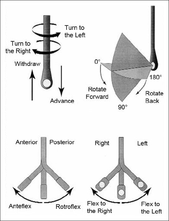
5 different ways you can physically manipulate the TEE probe

1. Withdraw or Advance up or down patient’s esophagus

2. Turn probe to right or left

3. Turn tip of flip in anterior- ante-flexing or in the posterior direction called retro-flexing --> large wheel

4. Turn tip to Left or right -->  small wheel (not typically used for our purposes)

5. In addition, you can rotate the transducer housed within the probe itself (AKA omniplane or multiplane)-->  adjusts the beam angle anywhere between 0° and 180° -->  two smaller buttons ( crystal rotation)

TEE manipulation.jpg


TEE-controls- wheels.png


The views are obtained in the following order: : 

The midesophageal 4-chamber view (ME 4C) is obtained by advancing the TEE probe to the thoracic esophagus and orienting the multiplane at 0-20° in neutral flexion. You may need to retroflex slightly to see all four chambers.

-   The midesophageal long-axis view (ME LAX) is obtained by leaving the probe in the same location as the midesophageal 4-chamber, but increasing the multiplane to between 110° and 160° while in neutral flexion. 

-   The transgastric short axis view (TG- SAX) is obtained by first moving the multiplane to 0°, then advancing the probe into the stomach and ante-flexing the probe

-   The bicaval view (ME bicaval) is obtained by turning the entire probe to the patient’s right towards the superior vena cava (SVC) and inferior vena cava (IVC) while in the mid-esophagus, keeping the multiplane at 90-100° with neutral flexion

 ( The first 3 views are recommended by ACEP. Bicaval not recommended by ACEP) 


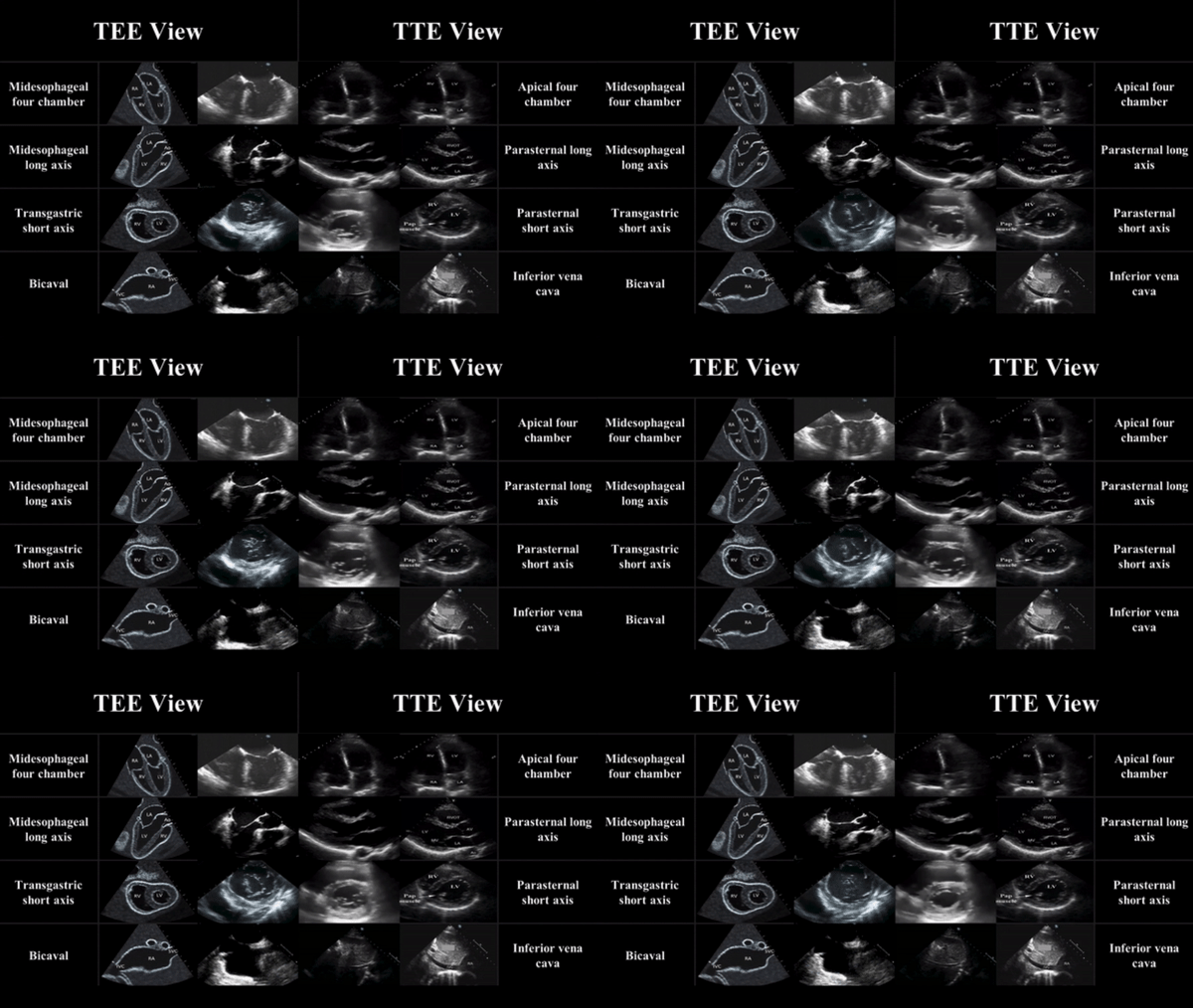
TEE views and their analogous TTE views


TTE-and-TEE.gif

Midesophageal four chamber view (ME 4C)

-  Apical four chamber view

-  Great visualization of all chambers as well as the tricuspid and mitral valves in one plane.

-   Evaluation of right and left ventricular systolic function and size

-   Preferred view to evaluate for the presence or absence of a perfusing rhythm during a pulse check.

ME4C.png


Midesophageal Aortic Long Axis view (ME LAX)

-  Midesophageal analogous to the parasternal long axis view in TTE

-   View includes the mitral and aortic valves, as well as the left atrium, left ventricle, and left ventricular outflow tract of the right ventricle.

-   Evaluate left ventricular systolic function, and provides feedback on compression adequacy and location. High-quality compressions cause maximal compression of the left ventricle and visualization of the aortic valve opening and closing indicating forward flow of blood.  Poor quality compressions are seen over the aortic root and there is no valvular indication of forward flow. 

ME- LAX.png


Transgastric Short Axis view (TG- SAX)

- Analogous to the parasternal short axis TTE view

- Evaluate left ventricular systolic function, including any regional wall motion abnormalities

- Can evaluate for acute MI and the presence of septal flattening in this view

TGSAX.png

Mid Esophageal Bicaval View (ME bicaval)

-   Analogous to the inferior vena cava view of TTE

-   Transducer plane cuts through the left atrium (LA), right atrium (RA), IVC and SVC.

This view allows the operator to evaluate for hypovolemia, atrial size, and interatrial septum bowing.

-   Aids in the placement of central venous catheters, transvenous pacemakers, or extracorporeal life support (ECMO) vascular cannulas by observing the initial wire placement in the vasculature

- Can aid inevaluation of fluid status to guide fluid resuscitation (looking at respiratory variation in SVC)

bicaval.png


Pitfalls

-  Compressions do not need to be stopped for TEE insertion. Additionally, the TEE can be left in the esophagus during defibrillation. The probe should be inserted or withdrawn while the tip is in neutral position, and not while the tip is flexed to avoid esophageal injury. 

-  Images should be optimized to avoid foreshortening of the ventricles and to include the appropriate structures for each view.

-  Pericardial effusions must be taken into clinical context, as small effusions can cause tamponade if accumulated rapidly, while large effusions can be well tolerated if they accumulate slowly.

-   Clotted hemopericardium may be isoechoic with the myocardium, making it difficult to identify.

-  Right ventricular failure is not specific to pulmonary embolism, and can be due to pulmonary hypertension or other etiologies such as right sided myocardial infarction, or even cardiac arrest itself.

-  Pleural effusions can be mistaken for pericardial effusions. Multiple views should be used to corroborate findings.

-  Fat pads can be mistaken for pericardial effusions, but these are hypoechoic rather than anechoic and limited to the anterior and apical regions of the heart, not circumferential.


 Resource: 

Check out this 3D module that you can practice on 

https://pie.med.utoronto.ca/TEE/TEE_content/TEE_standardViews_intro.html

References:

Drs Lawrence Haines, Judy Lin and Alyssa Phuoc-Ngyuyen

Images: Adapted from Arntfield R, Pace J, McLeod S, et al. Focused transesophageal echocardiography for emergency physicians-description and results from simulation training of a structured four-view examination. Crit Ultrasound J. 2015;7(1):27.

Teran, Felipe, et al. "Evaluation of out-of-hospital cardiac arrest using transesophageal echocardiography in the emergency department." Resuscitation 137 (2019): 140-147.

EmDocs

ACEP policy statement

https://www.acep.org/patient-care/policy-statements/guidelines-for-the-use-of-transesophageal-echocardiography-tee-in-the-ed-for-cardiac-arrest/

 ·