Electrophysiology 102: modes of pacing

Avid learners: 

As promised: part 2 on electrophysiology. The emphasis here will be modes of pacing and what they mean. Please note, I am not an electrophysiologist,

so if anything here is wrong, please respond ALL. 

This topic may require multiple reads, and I encourage you to seek out other sources. 

Please take this POD with a grain of salt.

Electrophysiology super-fellowships recently were extended to 2 years because this is a complicated topic.

We will focus on what the EP provider needs to know. 

Typically a pacing mode is described in letters. For simplicity sake, we will stick to the old fashioned 3 letters. 

A typical appearance is VVI [OO]. Most pacemakers will be reported with an omission of IV and V and will be just be the first 3 letters out of 5, in this case VVI. 

Position 1 is what is Paced

Position 2 is what is Sensed

Position 3 is what is the action of the device. 

Position 4 is rate modulation

Position 5 is for atrial, ventricular, or both pacing 

For this case (VVI), the ventricle is paced. The ventricle is also sensed, and if the pacemaker senses a ventricular beat, the pacemaker’s delivered beat will be inhibited.  So: if the rate of the pacemaker is set to 50 bpm, and the ventricle is beating at 60 bpm without pacing, every sensed beat actually inhibits the pacemaker from firing. However, if the ventricular rate all of sudden drops to 25, there are half the beats to sense, and the pacemaker will then start pacing. 

In short, for patients with an old school unipolar lead in the RV (VVI), if the ventricle does not beat faster than the programmed rate, the transvenous pacer will depolarize the RV. As I mentioned in my previous POD (Electrophysiology 101), this is why we see a LBBB morphology. 

Here is a key:  

miller_pacing_schema.png

reproduced from above source.

As per my review of the literature; the most confusing thing about pacing schema is the 3rd position: 

If a pacemaker is in DDD mode (most common mode), it will pace both chambers (if necessary) AND senses both chambers. So, if the atria does not spontaneously fire at the programmed rate, it will trigger an electrode signal causing pacing the atria; and subsequently, it will do the same for the ventricle. If an atrial depolarization occurs spontaneously, it will ensure there is a subsequent atrial beat. In short, DDD mode automatically senses which chamber(s) is/are not firing and makes them contract in synchrony. 

If a pacemaker is in DDI mode, “AV synchrony is provided only when the atrial chamber is paced.” Basically, this means the if the atrial chamber is not being paced because it is depolarizing on its own, the pacemaker will not depolarize the ventricle(s). But, if the atrial chamber needs to be paced, it will also subsequently pace the ventricle.

As mentioned previously in my POD, Cardiac Synchronization Therapy (CRT) expands the pacing to both ventricles in an attempt to improve cardiac output in the setting of reduced ejection fraction. 

By far the most important mode of pacing that we utilize emergently in the ED is VOO. In this mode we just overdrive pace the ventricle. Typically these are patients with bradycardia and/or hypotension/AMS. Occasionally, you may see intermittent asystolic events due to profound heart block. Both of these patients need to have transvenous pacing until electrophysiology can place a permanent pacemaker. You can let them figure out the mode that best suits them. 

Penultimate final point for patients with a pacemaker who come in unstable: if you a put a magnet on the pacemaker it will default the pacemaker to AOO, VOO, or DOO (all overdrive pacing). No response to this means dead battery or lead displacement, which means they need a revision. 

Final point: as I mentioned in my last POD on electrophysiology, putting a magnet on a pacemaker is substantially different than putting a magnet on an AICD. 

Magnet on pacemaker=overdrive mode

Magnet on AICD=inhibition of defibrillator

 · 

Cardiac Biomarkers Review

Dear Lifelong Learners and Readers of the TR POD: 

During one of the meetings on my admin rotation, this interesting question arose:

should we also be ordering serum creatinine kinase muscle/brain (CKMB) level on our ACS rule out patients?

This derives from Cardiology’s concern that there are too many patients with elevated troponin with catheterizations that were not significant for

TRUE

acute symptomatic coronary artery stenosis. 

In the absence of a STEMI or STEMI equivalent, we rely on serum biomarkers to support our hypothesis that the patient has myocardial ischemia.

Over the recent history of medicine, biomarkers supporting the diagnosis of myocardial ischemia have become increasingly sensitive. Which is to say, if the biomarker is negative, the condition you are testing for is more likely to be absent.

As the sensitivity increases, the probability with you can safely say a disease is not present also increases because the number of false negatives for that particular test decreases. However, this comes at the cost of an increasing number of false positives. The main problem that is highlighted in the graph below is that as we increase sensitivity, we increase the number of false positives, assuming a normal distribution.  

PastedGraphic-4.png

For those of you who want to review biostatistics (especially interns); take a look:

https://lifeinthefastlane.com/ccc/diagnostic-tests-in-research/

Below, is a graph showing different rises of biomarkers during a proven myocardial infarction (MI). Note that AST is not included in this graph, but it is very similar to the total CK line as per my review of the literature. 

PastedGraphic-3.png

As emergency providers, we are trying to rule out acute coronary syndrome (ACS) and suspicion for coronary artery disease (CAD). We aim to get into the nitty gritty of the statistics by which we practice: from both a historical perspective as well as a perspective of the clinician practicing today.

In the 50’s snd 60’s, the mainstay lab test was AST and CPK.

Sensitivity: 35%

Specificity: 58%

http://stenosis.heiderlab.de/

The above link offers a clinically derived calculator (non-validated) to predict the severity of stenosis. Presumably, the higher the AST, the higher the stenosis. 

So back in the day: if a patient had typical chest pain symptoms and elevate AST/CPK, they may have gone to the cath lab. Today, that would never fly, but back then it was all they had. 

In the 70’s doctors used the slightly more sensitive CKMB. 

Sensitivity: 65%

Specificity: 46%

In the 90’s there was low sensitivity cardiac-troponin which was used in combo with CKMB.

Sensitivity: 67%

Specificity: 74%

In the 2000’s, next generation cardiac troponin I and T assays were even more sensitive. 

Sensitivity: 89%

Specificity: 79%

***the reason for this low-ish sensitivity is due to the delay in elevation of troponin levels compared to other markers. The test is actually 99% sensitive when enough time has passed for the troponin to be elevated, given the appropriate clinical picture. 

Some institutions have

high sensitivity cardiac troponin

;

we don’t.

But FYI: Sensitivity is even higher and the specificity is lower. Because we don’t use this assay, we will not get into the specifics of this assay. 

Ironically, the newer “high sensitivity assays” that are available in Europe may decrease the false positive rate

. This is because there is higher resolution at the 99th percentile cut off point; which is to say: what we see as equivocal/borderline-positive troponin with our current assay may in fact be negative. Even though marketed as high sensitivity, that is actually not what the assay is in comparison to our current tests. What high sensitivity troponin means is more accuracy in defining non-zero troponin levels that are not positive for true ACS. 

Causes of elevated troponin that are not due to ACS as a result of critical CAD are broad: sepsis, stroke, pulmonary embolism, CHF, renal failure, myocarditis and pericarditis. 

So, a mild to moderately elevated troponin level in the setting of another disease process does not warrant a hard push to the cath lab unless it is indicated by the patient’s particular presentation. 

To come back full circle to the initial question:

is CKMB, myoglobin, or CK indicated?

The answer is complicated. It should be case specific. Any cardiac chest pain with recent onset (less than 4 hours) that is concerning for MI, I would add on myoglobin and CKMB. This could be especially important if you are dealing with unstable angina with intermittent chest pain. If the myoglobin is elevated and the patient has chest pain with a normal troponin and a non-ischemic appearing EKG, I would use that elevated myoglobin and active chest pain to get a prompt cardiology evaluation. In these cases, the troponin becomes elevated later. 

Moreover, our cardiology colleagues are seeking more data to push them one way or another in the absence of slam-dunk EKG findings. After reviewing the literature, in my opinion:

our troponin assay should be sufficient with serial testing

UNLESS

the patient has had recent MI, PCI, a comorbid condition that can elevate the troponin level, or

chest pain that started

minutes ago

. If they have a comorbid condition, CK, CKMB, and Myoglobin may help differentiate the cause of the elevated troponin. While none of these tests will be a gold standard like a cardiac catheterization, the results can certainly help our Cardiology colleagues who will undoubtedly be consulted as inpatients for “NSTEMI”. For example, an elevated troponin with a normal myoglobin might be

very reassuring. 

No test is perfect.

There is always some degree of uncertainty regarding the significance of a result. 

If they have chest pain and it is early but no STEMI or STEMI equivalents on EKG, get that CKMB and Myoglobin.

They’re cheap tests

. If you get a patient with an elevated myoglobin, non-ischemic EKG, but has active chest pain, that is one more argument that you have to advocate for a cardiac catheterization. 

TR,

W

References:

https://path.upmc.edu/cases/case178/dx.html

http://www.ijmedicine.com/index.php/ijam/article/viewFile/317/287

https://step1.medbullets.com/stats/101006/2x2-tables-sn-sp-ppv-npv-or-rr

https://www.slideshare.net/cksheng74/sensitivity-specificity-and-likelihood-ratios

Giannini EG, Testa R, Savarino V. Liver enzyme alteration: a guide for clinicians. CMAJ : Canadian Medical Association journal = journal de l'Association medicale canadienne. 2005; 172(3):367-79.

Lewandrowski KB. Cardiac markers of myocardial necrosis: a history and discussion of milestones and emerging new trends. Clinics in laboratory medicine. 2014; 34(1):31-41, xi.

Mannu GS. The non-cardiac use and significance of cardiac troponins. Scottish medical journal. 2014; 59(3):172-8.

Sethi A, Bajaj A, Malhotra G, Arora RR, Khosla S. Diagnostic accuracy of sensitive or high-sensitive troponin on presentation for myocardial infarction: a meta-analysis and systematic review. Vascular health and risk management. 2014; 10:435-50. [pubmed]

 · 

Electrophysiology 101

One of my favorite topics that is as nerdy (if not more so) than toxicology is a subspecialty of cardiology called electrophysiology. While I am not an expert at any field at this point, I certainly enjoyed learning from our prestigious faculty in the CICU. This is the first of a 2 part pearl of the day. The next pearl of the day, we will get into the various modes of pacemaking and what they mean.

So let us review: pacemakers, AICD’s, the different varieties, why people have them, and what we as ED providers need to know.

Electrical Conduction system of the heart

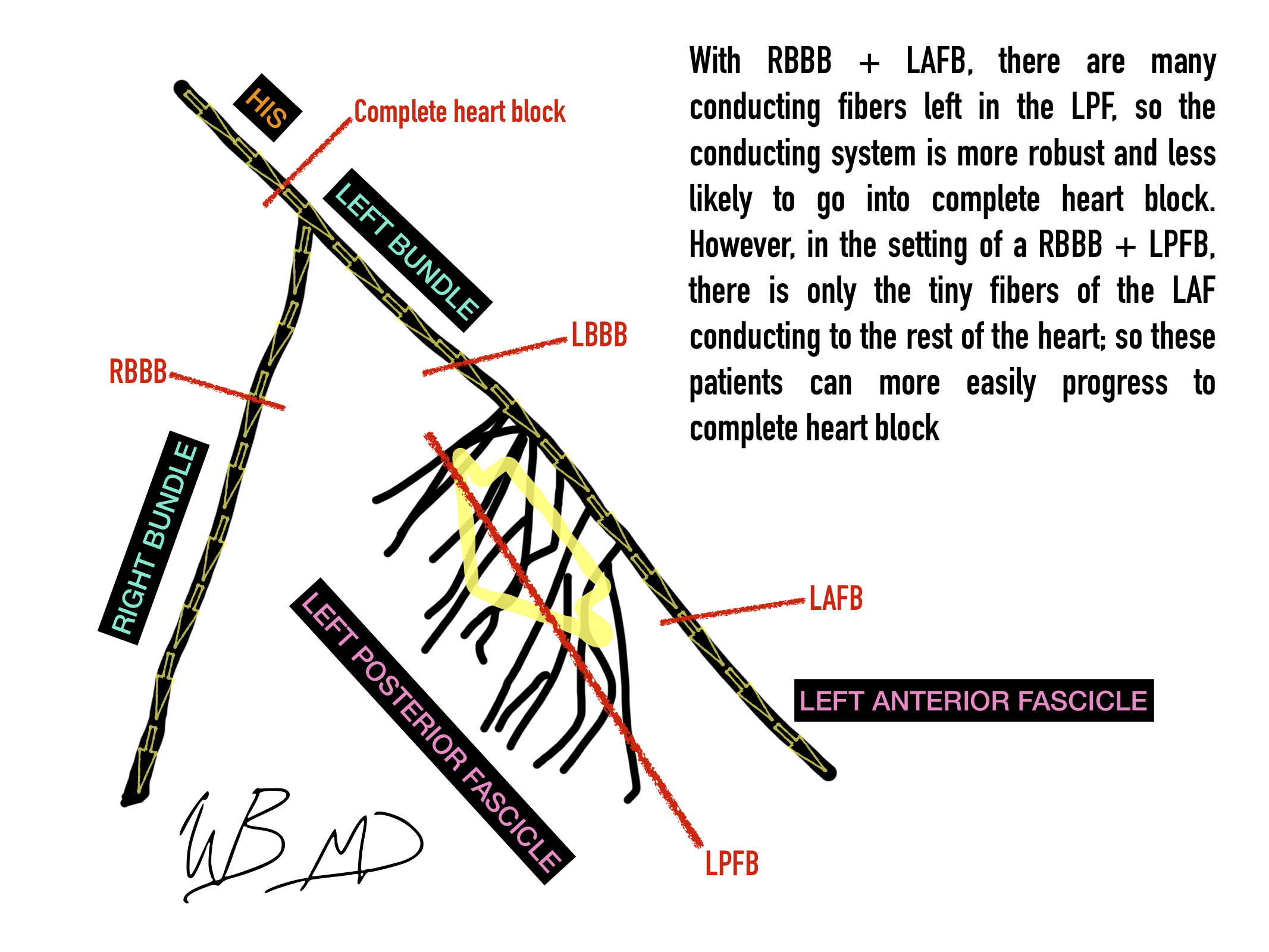
I have attached an infographic showing the various points of heart block. Notice how the left posterior fascicle has many conducting fibers. This important, because in the setting of RBBB + LAFB, there are many conducting fibers left so the conducting system is more robust and unlikely to go into complete heartblock. However, in the setting of a RBBB + LPFB, there is only the tiny fibers of the LAF conducting to the rest of the heart; so these patients need a pacemaker.

electrical-conducting-system-of-the-heart-text.jpg

Pacemakers

Complete heartblock or ecg findings that they will likely soon develop complete heart block (eg mobitz type 2 and combined RBBB with left posterior fascicular block) are indications for pacemaker placement. If they are unstable on presentation, they need an emergently placed transvenous pacer.

A single lead pacemaker tunnels through the venous system and delivers electrical impulses to the right ventricle. The wave of depolarization starts in the right ventricle; thus, the ECG finding of a paced rhythm is of a widened QRS seen in a LBBB.

For patients with reduced EF’s who need pacing, there is a current trend to place biventricular pacemakers. With these devices, a lead is placed into both the right and left chamber of the heart. This is termed cardiac resynchronization therapy (CRT). Because both the chambers of the heart are paced simultaneously, the QRS narrows and it is thought this improves EF.

Automatic implantable Cardiac Defibrillators (AICD’s)

There are various models of AICD’s and are capable of providing single chamber pacing and CRT in all of the various modes depending on model. There main function is to monitor the heart for dangerous arrhythmia and deliver electrify to abort the dangerous rhythm (usually tach or vfib).

Aside from there pacemaking and/or automatic defibrillator function, all of these devices are essentially continuous cardiac monitors. As such, any patient who has one of these devices who presents to our ED can have there device interrogated.

Mostly for the interns: any syncope, report of AICD discharge, palpitations, etc with an implantable device should have there device interrogated.

Special note: placing a magnet on an AICD will turn off the defibrillator mode. This should be performed after a failed resuscitation or in a DNR/DNI patient at the point of death.

 ·