POD Aortic Dissection

A patient came to the north side today with an acute aortic dissection. Here are images obtained by the ultrasound team when the patient first came in.

A suprasternal view showing an intimal flap:

suprasternal.jpg

A short axis view of the abdominal aorta showing an intimal flap

abdominal.jpg

Diagnosis was made, BP meds started, cardiothoracic consulted, and CT expedited.

CT showed a severe type B thoracoabdominal aortic dissection:

CT-aorta.jpg

Aortic Dissection

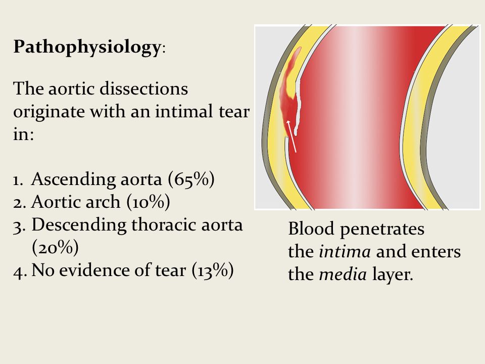
Pathophysiology:

Tear in the intima (inner most layer), bleeding into the media (middle layer)

Pathophysiology_Theaorticdissectionsoriginatewithanintimaltearin_Ascendingaorta65Aorticarch10.jpg

Diagnosis of aortic dissection is very time sensitive:

mortality is directly proportional to time elapsed between symptom onset and diagnosis/treatment

.

How does it eventually kill you? (I think it’s important to ask this question about all disease processes)

acute aortic regurgitation —> cardiogenic shock

Cardiac tamponade —> obstructive shock

Major brach-vessel obstruction —> vasodilatory shock from dead organ or limb

Aortic rupture —> hemorrhagic shock

2 types that we care about: Stanford Type A and Stanford Type B

types.jpg

Type A

:

involves ascending aorta

— surgical — a/w aortic rupture, tamponade, aortic regurg, AMI, stroke — more common (68%)

Type B

:

does not involve ascending aorta — medical (BP control and monitoring) — a/w limb/organ ischemia  — less common, (32%) — usually originates just distal to L subclavian artery

Classic history: old person,

very hypertensive

;

abrupt onset

,

tearing/ripping chest pain

,

radiating to bac

k; a/w neuro symptoms e.g.

weakness/numbness

(due to vessel branch occlusion); a/w syncope/diaphoresis/N/V

Other risk factors include Marfan’s, connective tissue disease, FHx aortic disease, known aortic valve disease, recent aortic manipulation (e.g. TAVR, surgery), known thoracic aortic aneurysm, tobacco;  rarely 3rd trimester pregnancy, TB, syphilis,  vasculitis, blunt trauma

Classic physical: Pulse deficit (present in <20% of cases), unequal BP in upper/lower extremities, neuro deficits, signs of tamponade

Diagnosis:

Labs: basics, coags, trop, consider d-dimer (actually high sensitivity/NPV for dissection due to blood often clotting I false lumen)

CT angio aorta: gold standard for diagnosis of aortic dissection

CXR: not sensitive, not specific — sometimes mediastinal or aortic knob widening, few other nonspecific signs

TEE: is an excellent modality that’s in the works but we don’t have it operational yet

TTE: next best thing, as usual with ultrasound it’s specific but not sensitive - see below

Ultrasound for aortic dissection — obtain the following views:

Subxiphoid: look for pericardial effusion
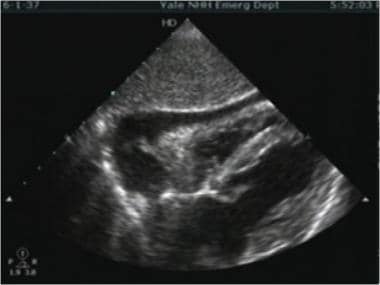
Image result for subxiphoid effusion'

Parasternal long: look for effusion, look at the descending aorta, look for aortic regurg with color doppler and measure the aortic root (nl <4cm) if you want to be fancy

para-long.jpg

Suprasternal window:

look for dissection flap (image from University of Maryland department of cardiology)

Probe above the the patient’s sternum pointed inferoposteriorly with probe marker to patient’s left (assuming cardiology convention)

SSNV.jpg

Abdominal aorta scan: look for dissection flap from diaphragm to iliacs, also measure diameter in short and long

Management (From the AAC/AHA aortic dissection guidelines):

ACC AHA AoD Treatment-Algorithm

Note: When blood pressure is intact, first bring heart rate with beta blockers, then control pain, then see if they need further BP control.

 · 

Friday Pearl of the Day: Not Your Ordinary STEMI Notification

We’re going to go though this POD as a case rather than me yelling facts at you. READ ON!

Red phone rings. “We’re bringing in a STEMI. Vitals are HR 155, BP 75/55, RR 35, 90% on NRB. Our prehospital EKG should be in the email system already.” Cath lab fellow is notified.

pasted-image.jpeg

The patient is brought into resus. Middle aged man, diaphoretic, holding his chest, tachypneic but no accessory muscle use. “Help me doc. I think I’m dying. My chest hurts so bad and I can’t breathe… What’s that? No I don’t take meds or have medical problems but I don’t really see doctors either.”

You listen to his lungs: crackles b/l; ultrasound his lungs: B-lines b/l… seems like acute pulmonary edema but he’s hypotensive so nitro’s out of the question. Furthermore, looks like it could be RV MI. You call for BiPAP (cautiously because that could make him more hypotensive too), which he tolerates and he feels better. A small dose of fentanyl helps with his pain as well.

Meanwhile, cardiology is busy consenting him for cath lab, you put in orders, and labs are being drawn. Another EKG and set of vitals are repeated, essentially the same as prehospital.

Chest X-ray shows up and shoots an image.

pasted-image-2.jpeg

Oh damn, this guy really does have pulmonary edema. Why does this guy have acute pulmonary edema? You astutely go back and put the probe on his chest again to look at the heart.

His parasternal window is garbage but his apical window is decent. Below is your apical long (same probe orientation as parasternal long but in the apical position).

pasted-image-3.jpeg

...there’s something flapping around on his mitral valve… it’s his papillary muscle! This guy ruptured his papillary muscle!

Papillary muscle rupture

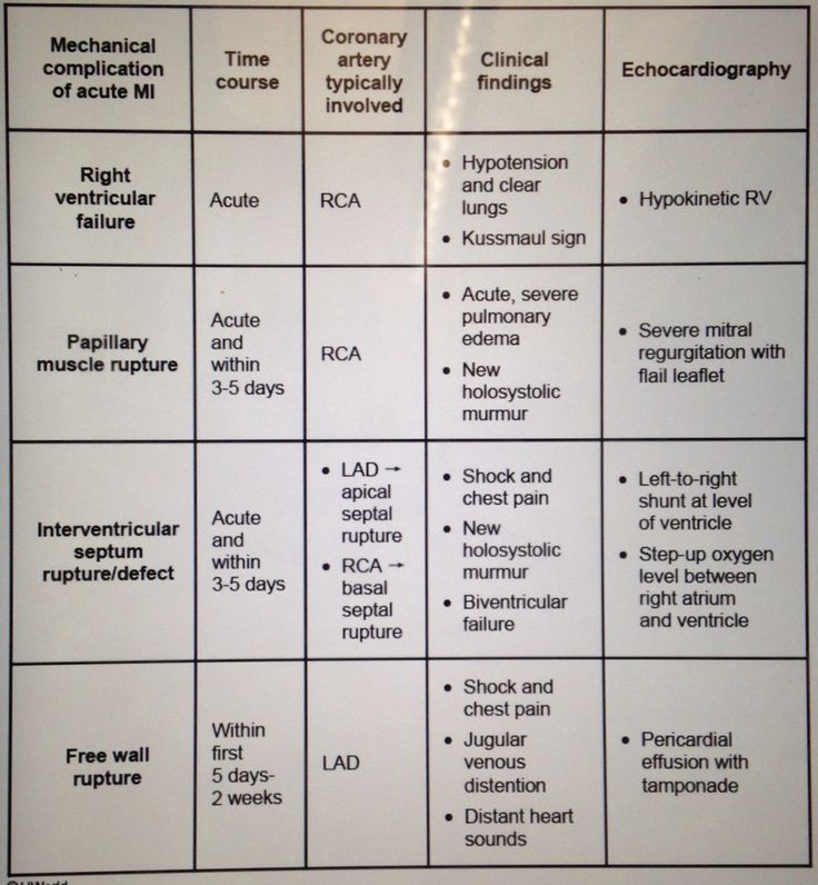
… you have a sudden flashback to studying for USMLE Step 1. A table that you once studied comes to your mind…

pasted-image-6-1.jpeg

Right! It’s one of the

mechanical complications of acute MI

… happens with

RCA infarcts

, can happen

acutely and within 3-5 days

acute severe pulmonary edema with flail leaflet on the echo

... bad news…

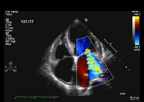
Your flashback ends and you’re back in the ED ultrasounding this guy. His mitral regurgitation is pretty insane when you put some color on it.

pasted-image-4.jpeg

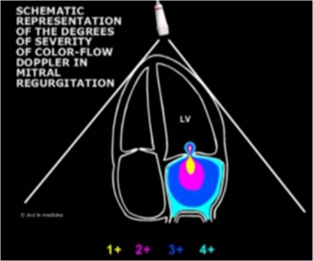
Probably 4+

pasted-image-5.jpeg

Suffice it to say, cardiology doesn’t want to take him to the cath lab any more, but at least the fellow documents an official echo to back you up. You change your underwear and

call cardiothoracic surgery

. They’re booking him for the OR immediately to repair his valve.

As an academic exercise, you and the cardiology fellow take a second to listen to his murmur. God forbid you had to diagnose this without ultrasound.

pasted-image.png

There it is,

holosystolic at the apex

.

He’s still hypotensive though and now he’s getting a little altered. The OR isn’t ready yet.

If he wasn’t hypotensive you could use nitroprusside to reduce his afterload

and improve his forward cardiac output… but since he’s hypotensive, cardiology helps you set up the only

other option: an intra-aortic balloon pump

to support perfusion to his heart and brain until surgery.

34982840_372122586646272_1955625577692004352_n.jpg

Off to the OR he goes. His operative mortality is 50% but it’s his best shot. With medical therapy alone, his mortality would be 75% at 24 hours and 95% at 2 weeks.

Well done, doc. He has a fighting chance now.

 · 

SGARBOSSA'S CRITERIA POD

SGARBOSSA CRITERIA

  • Used to diagnose a myocardial infarction in an ECG with LBBB or ventricular paced rhythm

  • Score ≥3 90% specific for acute MI but only 36% sensitive.

    • Low score cannot rule out MI.

Original Criteria: less sensitive in detecting MI

  • Concordant ST elevation > 1mm in leads with a + QRS complex (+5 points)

  • Concordant ST depression > 1mm in V1-V3 (+3 points)

  • Excessively discordant ST elevations > 5mm in leads with a negative QRS complex (+ 2 points)

Modified Criteria: more sensitive in detecting myocardial infarction

Eliminates the point system.

  • ≥ 1 lead with ≥1 mm of concordant ST elevation

  • ≥ 1 lead of V1-V3 with ≥ 1 mm of concordant ST depression

  • ≥ 1 lead anywhere with ≥ 1 mm STE and proportionally excessive discordant STE, as defined by ≥ 25% of the depth of the preceding S-wave

 ·