Palliative Dyspnea

Managing dyspnea in the palliative patient.

This comes down to 4 approaches:

  • Oxygen

  • Opiates

  • Benzodiazepenes

  • Addressing the underlying issue

  • Other measures of comfort

Oxygen

  • Several options here with pro's and con's to all

  • Nasal Cannula 

    • Comfortable at low flows

    • Limited in how much oxygen it can deliver as it provides no reservoir of oxygen; it depends on the patient's upper airway as the reservoir of oxygen

    • at high flow rates is uncomfortable and causes dryness and bleeding unless delivered with a humidifier)

    • Many patients mouth breathe at the end of life

  • Non-rebreather

    • provides more oxygen, enables oxygen delivery to mouth breathers

    • Uncomfortably noisy, must be drawn tightly against the face to be most effective

    • muffles communication at a time when it is of key importance in the dying patient

    • Dries patient's mouth and nares out

  • Venturi Mask

    • An underutilized therapy

    • Addresses mouth breathing

    • Mixes oxygen with room air

    • Able to provide relatively high flow rates of oxygen 

    • Does not need to be humidified as high flow rates of oxygen are mixed with ambient room air

  • High-flow nasal cannula

    • Comfortably provides humidified oxygen at extremely high rates

    • Does not provide oxygen to mouth breathers

    • If the patient is being admitted it requires admission to the MICU (or potentially PAMCU)

  • Non invasive ventilation (Bipap)

    • Noisy, uncomfortable, frightening

    • Decreases the ability to commmunicate

Opioids

  • THE KEY TO PALLIATIVE DYSPNEA

  • Can be delivered via the subcutaneous route, another underutilized therapy

  • Administer zofran to offset possible associated nausea

  • Decrease the intensity of air hunger and dyspnea related anxiety

  • Have been shown to NOT SHORTEN LIFE IN PALLIATIVE PATIENTS, which is important to communicate to the dying patient's family. 

Benzodiazepenes

  • Anxiety leads to worsening dyspnea; managing the anxiety therefore aids in management of dyspnea

  • Generally not used as monotherapy, however can be used in addition with opiates in the anxious and dyspneic patient

Other measures

  • Position the patient as they wish, though generally the more upright patient is the more comfortable patient

  • Death rattle: As patients lose consciousness they lose their ability to swallow and oral secretions can pool, causing gurgling noises. There is no evidence that this is disturbing to patients, but families often have a very hard time with these noises.

    • Glycopyrrolate can help mitigate this disturbing noise

Cause specific techniques = address the underlying issue

  • Must weigh the benefits vs. the discomfort of performing these interventions

  • Pleural effusions: Thoracentesis

  • Anemia: Transfusion

  • Obstructing airway mass: Steroids, palliative radiation if available

  • Pneumonia: Antibiotics

  • Fluid overload: Diuresis

  • Bronchospasm: Bronchodilators

See:

https://first10em.com/palliative-resuscitation-dyspnea/

https://www.rtmagazine.com/products-treatment/monitoring-treatment/therapy-devices/oxygen-administration-best-choice/

 · 

Treating Laryngospasm

LARYNGOSPASM
You’re doing a procedural sedation in pediatrics. Despite your attending’s forewarning, you push that IV ketamine a bit too quickly. Suddenly, you hear a loud “crowing” or “squeaking” sound and look up at the monitor to see a flat line on capnography. Your heart sinks as it dawns upon you that you caused the much-dreaded laryngospasm.

WHAT DO I DO?!
1. Stop all procedures
2. Perform Larson’s maneuver - this is a modified jaw thrust maneuver where pressure is applied towards the top of the ramus of the mandible

laryngospasm-notch.jpg

3. Use a bag valve mask with PEEP valve and 100% oxygen to provide continuous positive airway pressure
4. Ask your team to prepare for intubation
5. Deepen anesthesia with IV propofol (0.5mg/kg IV push)

Most cases of laryngospasm will resolve with these maneuvers and propofol. In the rare event it doesn’t…

6. Give an IV paralytic (rocuronium 1mg/kg IV or succinylcholine 1.5mg/kg IV) and proceed to intubate.

Sources: Justin Morgenstern, "Managing laryngospasm in the emergency department", First10EM blog, March 3, 2016. Available at: https://first10em.com/laryngospasm/.

Alejandro Romero

EM PGY-3


 · 

Trauma in Pregnancy

Resuscitation of the Pregnant Trauma patient


General principles

·      Trauma is the most common cause of non-obstetrical maternal death in the United States

·      Best fetal resuscitation is good maternal resuscitation.

·      Stabilization of the pregnant women is the first priority; then, if the fetus is viable (≥ 23 weeks), fetal heart rate auscultation and fetal monitoring can be initiated and an obstetrical consultation obtained as soon as feasible

·      In Rh-negative pregnant trauma patients, quantification of maternal–fetal hemorrhage by tests such as Kleihauer-Betke should be done to determine the need for additional doses of anti-D immunoglobulin.

·      Tetanus vaccination is safe in pregnancy and should be given when indicated.


Airway

·      Greater risk for difficult intubation than non-pregnant patient

·      Pregnancy related changes à decreased functional residual capacity, reduced respiratory system compliance, increased airway resistance, and increased oxygen requirements

·      Gastric emptying is delayed in pregnancy à greater risk for aspiration

·      Respiratory tract mucosal edema à A smaller size of endotracheal tube is recommended

·      Choice of RSI medications NOT affected by pregnancy status


Breathing

·      Place chest tube one to 2 intercostal spaces higher than usual to account for displacement of the diaphragm during pregnancy

·      Marked increases in basal oxygen consumption à lower threshold for supplemental oxygen


Circulation

·      Fluid and Colloid resuscitation like standard trauma protocol

·      Uteroplacental vasculature is highly responsive to vasopressors, and their administration may decrease placental perfusion à vasopressors should be avoided unless refractory

·      Avoid supine hypotension: Compression of IVC by the uterus can cause up to 30% reduction in cardiac output à Place in left lateral position or by manual displacement of the uterus while the injured patient is secured in the supine position

·      O-negative blood should be transfused in order to avoid Rh sensitization in Rh-negative women

·      Vital signs: heart rate increases by 15% during pregnancy. Tachycardia and hypotension, typical of hypovolemic shock, may appear late in the pregnant trauma patient because of her increased blood volume.

·      Maternal vital signs and perfusion may be preserved at the expense of uteroplacental perfusion, delaying the occurrence of signs of hypovolemic shock

·      Attempt to obtain supra-diaphragmatic intravenous or intraosseous access for volume resuscitation and medication administration.


FAST

·      The FAST is less sensitive for free fluid in the pregnant patient than in non-pregnant patients.  Sensitivity decreases with increasing gestational age, likely due to altered fluid flow within the abdomen.

·      Management of suspected placental abruption should not be delayed pending confirmation by ultrasonography as ultrasound is not a sensitive tool for its diagnosis.


Secondary survey

·      In cases of vaginal bleeding at or after 23 weeks, speculum or digital vaginal examination should be deferred until placenta previa is excluded by a prior or current ultrasound scan.


Imaging

·      Radiographic studies indicated for maternal evaluation including abdominal computed tomography should not be deferred or delayed due to concerns regarding fetal exposure to radiation.

·      Ionizing radiation has the highest teratogenic potential during the period of organogenesis (5–10 weeks), with an increased risk of miscarriage before this period.

·      With abdominal CT during the third trimester the fetal exposure is around 3.5 rads, which is still under the threshold for fetal damage

·      Contrast agents should be used if indicated.


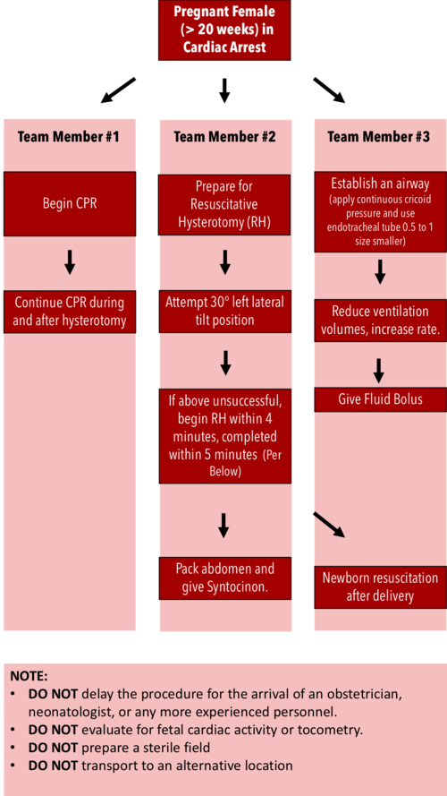
Resuscitative Hysterotomy in Cardiac Arrest

·      Should begin within 4 minutes and completed within 5 minutes of cardiac arrest

·      Both maternal and fetal survival decrease significantly after 5 minutes

·      Do NOT delay the procedure for the arrival of an obstetrician or neonatologist.

·      Do NOT evaluate for fetal cardiac activity or tocometry.

·      Do NOT prepare a sterile field (but be as clean as possible).

·      Do NOT transport to an alternative location.

·      Performing RH increases maternal cardiac output by 30%.


RH Algorithm.png


References:


Tamingthesru.com

EmDocs

Jain, Venu, et al. "Guidelines for the management of a pregnant trauma patient." Journal of Obstetrics and Gynaecology Canada 37.6 (2015): 553-571.

Smith, Kurt A., and Suzanne Bryce. "Trauma in the pregnant patient: an evidence-based approach to management." Emergency medicine practice 15.4 (2013): 1-18.


 ·