Trauma in Pregnancy

Resuscitation of the Pregnant Trauma patient


General principles

·      Trauma is the most common cause of non-obstetrical maternal death in the United States

·      Best fetal resuscitation is good maternal resuscitation.

·      Stabilization of the pregnant women is the first priority; then, if the fetus is viable (≥ 23 weeks), fetal heart rate auscultation and fetal monitoring can be initiated and an obstetrical consultation obtained as soon as feasible

·      In Rh-negative pregnant trauma patients, quantification of maternal–fetal hemorrhage by tests such as Kleihauer-Betke should be done to determine the need for additional doses of anti-D immunoglobulin.

·      Tetanus vaccination is safe in pregnancy and should be given when indicated.


Airway

·      Greater risk for difficult intubation than non-pregnant patient

·      Pregnancy related changes à decreased functional residual capacity, reduced respiratory system compliance, increased airway resistance, and increased oxygen requirements

·      Gastric emptying is delayed in pregnancy à greater risk for aspiration

·      Respiratory tract mucosal edema à A smaller size of endotracheal tube is recommended

·      Choice of RSI medications NOT affected by pregnancy status


Breathing

·      Place chest tube one to 2 intercostal spaces higher than usual to account for displacement of the diaphragm during pregnancy

·      Marked increases in basal oxygen consumption à lower threshold for supplemental oxygen


Circulation

·      Fluid and Colloid resuscitation like standard trauma protocol

·      Uteroplacental vasculature is highly responsive to vasopressors, and their administration may decrease placental perfusion à vasopressors should be avoided unless refractory

·      Avoid supine hypotension: Compression of IVC by the uterus can cause up to 30% reduction in cardiac output à Place in left lateral position or by manual displacement of the uterus while the injured patient is secured in the supine position

·      O-negative blood should be transfused in order to avoid Rh sensitization in Rh-negative women

·      Vital signs: heart rate increases by 15% during pregnancy. Tachycardia and hypotension, typical of hypovolemic shock, may appear late in the pregnant trauma patient because of her increased blood volume.

·      Maternal vital signs and perfusion may be preserved at the expense of uteroplacental perfusion, delaying the occurrence of signs of hypovolemic shock

·      Attempt to obtain supra-diaphragmatic intravenous or intraosseous access for volume resuscitation and medication administration.


FAST

·      The FAST is less sensitive for free fluid in the pregnant patient than in non-pregnant patients.  Sensitivity decreases with increasing gestational age, likely due to altered fluid flow within the abdomen.

·      Management of suspected placental abruption should not be delayed pending confirmation by ultrasonography as ultrasound is not a sensitive tool for its diagnosis.


Secondary survey

·      In cases of vaginal bleeding at or after 23 weeks, speculum or digital vaginal examination should be deferred until placenta previa is excluded by a prior or current ultrasound scan.


Imaging

·      Radiographic studies indicated for maternal evaluation including abdominal computed tomography should not be deferred or delayed due to concerns regarding fetal exposure to radiation.

·      Ionizing radiation has the highest teratogenic potential during the period of organogenesis (5–10 weeks), with an increased risk of miscarriage before this period.

·      With abdominal CT during the third trimester the fetal exposure is around 3.5 rads, which is still under the threshold for fetal damage

·      Contrast agents should be used if indicated.


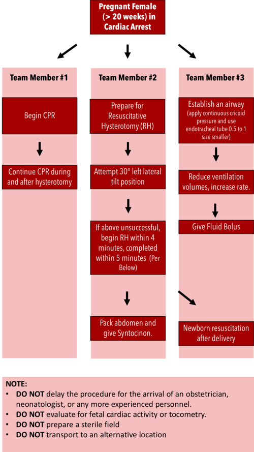
Resuscitative Hysterotomy in Cardiac Arrest

·      Should begin within 4 minutes and completed within 5 minutes of cardiac arrest

·      Both maternal and fetal survival decrease significantly after 5 minutes

·      Do NOT delay the procedure for the arrival of an obstetrician or neonatologist.

·      Do NOT evaluate for fetal cardiac activity or tocometry.

·      Do NOT prepare a sterile field (but be as clean as possible).

·      Do NOT transport to an alternative location.

·      Performing RH increases maternal cardiac output by 30%.


RH Algorithm.png


References:


Tamingthesru.com

EmDocs

Jain, Venu, et al. "Guidelines for the management of a pregnant trauma patient." Journal of Obstetrics and Gynaecology Canada 37.6 (2015): 553-571.

Smith, Kurt A., and Suzanne Bryce. "Trauma in the pregnant patient: an evidence-based approach to management." Emergency medicine practice 15.4 (2013): 1-18.


 · 

Holy smokes! Inhalation Injury

First steps (stable pt)
-treat as a trauma pt (ABCDE) and look for traumatic injuries
-place pt on NRB with O2 to 15 L/m
—add nebs 4% lidocaine early to prepare for visualization of cords with videolargynoscope
4:2:1 rule for burn pt fluid resuscitation
—give fluids even if no external burns visible, as pt will have insensible losses
-treat pain!
-evaluate cords and surrounding laryngeal structures for edema with video laryngoscope or bronch

How to risk-stratify your patient with suspected smoke inhalation injury:

Screen Shot 2018-12-27 at 12.04.46 PM.png

Rule out
carbon monoxide toxicity: obtain serial blood gases (send co-oximetry) to monitor carboxyHb. Normal levels are 5 – 12%, depending on whether or not the pt is a smoker. Also consider if family presents with similar vague symptoms.
-cyanide toxicity: cyanide levels are not reliable in excluding toxicity, as it is rapidly cleared, and don’t result for days. Use lactate>8 or rising lactate despite fluid resuscitation to raise suspicion for toxicity
—ddx for elevated lactate (=impaired tissue oxygenation) in burn pt: cyanide, metHb, hypoxia, volume depletion
-look for rhabdo and AKI

Warning signs of respiratory failure
drooling or difficulty swallowing = impending failure
-monitor for stridor, hoarseness, and respiratory distress
-PaO2/FiO2 ratio indicates degree of pulmonary shunting past injured lung. PaO2/FiO2<300 forewarns respiratory failure

Intubation
-early elective intubation in a controlled setting is better than crash intubation of a pt with edematous airway structures
-Prepare multiple sizes ETTs in anticipation of vocal cord edema. Use the largest that will fit so that the pt can get a bronchoscopy upstairs. Prepare suction for soot-filled secretions. Sux is safe to use up to 24h post-burn.
-use volume controlled ARDS settings (6-8 mL/kg TV)
—airways and lung become less compliant in inhalational injury, so must prevent barotrauma and allow for permissive hypercapnea

 · 

The Bougie POD

Awwww yisss, airway stuff!

I’d like to start this POD off by talking about the study that got everyone buzzing about the bougie this summer, published last June in JAMA by Driver et al. at Hennepin:

Effect of Use of a Bougie vs Endotracheal Tube and Stylet on First-Attempt Intubation Success Among Patients With Difficult Airways Undergoing Emergency Intubation, A Randomized Clinical Trial

Numbers and outcomes:

  • They randomized 757 patients: 381 to a bougie-first approach, 376 to a traditional styletted ETT-first approach

  • Population was >18, undergoing intubation with a Macintosh (standard geometry) blade (direct or video, how much the intubator looked at the screen was at the teams discretion)

  • 380 patients had at least one difficult airway feature

  • Success on 1st attempt was 98% in bougie-first group vs 87% in ETT+stylet group, P=0.0001, NNT=9

  • Success on 1st attempt in patients with difficult airway features was 96% in bougie-first group vs 82% in ETT+stylet group, P<0.0001, NNT=7

Let’s just state what that last NNT means in words to let it sink in: You have use a bougie on 7 patients with difficult airway features in order to prevent one first-pass failure.

Furthermore, the bougie held its own among every stratification, e.g.:

  • Obese patients: (96% vs 75%)

  • Patients that needed cervical in-line stabilization: (100% vs 78%,)

  • Patients with poor views (Cormack-Lehane grades 2 to 4): (97% vs 60%)

A few other noteworthy things:

  • The duration of the first pass was about the same between bougie and ETT groups,

  • The total time of intubation was far longer in the ETT group, owing to more often needing multiple attempts passes

  • No difference in complication rate or direct airway trauma

Bottom line: This is extremely compelling evidence that first pass success is improved with use of a bougie.

We massively underutilize the bougie. Let’s improve our first pass success and use it more often.

I would especially consider using a bougie as first pass if you’re a less-experienced intubator or you’re starting to learn DL. Furthermore, even if you want to be old school and use it “only as a backup/rescue device”, heaven help you if you actually have to use it as such and have never practiced using it.

For anyone that may not be 100% familiar…

How to use a bougie:

  • get a view

  • pass your bougie through the cords, the coudé tip helps guide it anteriorly where it needs to go

  • you know you’re in the trachea because it stops around the carina (be gentle, airway perforations are sub-optimal), you can also theoretically feel the subtle clicking of the tracheal rings as it slides down the trachea

  • your assistant slides the tube over the back end of the bougie and then stabilizes the back of the bougie while you railroad the tube over it and through the cords

  • keep retracting the tongue with the laryngoscope while you do this to facilitate passage

  • you may encounter some resistance when it reaches the arytenoids; twisting the tube solves this problem

  • you can definitely do all this by yourself too, it’s just a little trickier to maneuver all the moving parts

The bougies used in the Hennepin study were 70 cm gum elastic (blue) bougies, the same ones we stock in our ED. These as well as slightly shorter 60cm bougies are stocked in most departments you might work in. Thanks to Reuben we also now stock the purple malleable bougies! These excellent devices and will save you when you run into weird geometry and can even be used with hyperangulated laryngoscopes but this is a little harder. When I use them as a regular bougie, I’ll usually give them a slight coudé tip and mild anterior bend like that of the ETT and revise if necessary. The stopper can be taken off or used to pre-load the tube.

 ·