HPI
29M with no PMH presents to the ED with worsening RLQ pain since last night. He endorses subjective fever, and ROS is otherwise negative. On exam, he has abdominal tenderness to palpation with voluntary guarding.
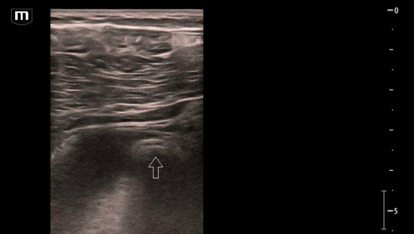
Bedside ultrasound of the RLQ demonstrates a dilated, tubular, blind-ending structure measuring 17mm. An appendecolith is noted. Findings consistent with acute appendicitis.
CT abdomen/pelvis confirmed the diagnosis of acute appendicitis, with an appendecolith and trace free fluid, for which perforation cannot be excluded.
Pearls
For most pediatric patients, choose the linear probe, but can switch to the curvilinear probe for larger patients
Place the probe on McBurney’s point or the point of maximal pain and use a lawnmower technique to scan the area
Anatomical landmarks: iliac crest, iliac artery, psoas muscle
Signs of appendicitis on ultrasound
A non-compressible tubular structure > 6 mm (append-“six") in diameter
Sometimes a fecalith (appendecolith) can be seen with posterior shadowing
Secondary signs include hyperechoic “hot fat”, free fluid, hyperemia, and bowel wall edema
Case Conclusion
Patient underwent a successful laparoscopic appendectomy.
Here is a helpful Five Minute Sono video about appendicitis: https://coreultrasound.com/appendicitis/
Happy scanning!


