94 yoF with PMHx of gastric cancer and recent SBO (managed non-operatively) presented to the ED with worsening abdominal pain, constipation, and obstipation.
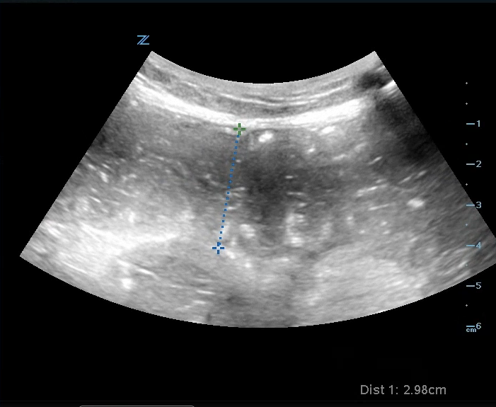
An ultrasound was performed that showed multiple signs consistent with an SBO:
Image 1: Dilated loops of bowel > 2.5 cm.
Video 1: To-and-for movement of fluid in the bowel. Normally, feculent material should only move in the direction of peristalsis. However, if there is a distal obstruction, you will see feces move back and forth as it attempts to move past it.
Video 1: Keyboard sign - when the plicae circulares, finger-like projections of the jejunal inner wall, become more prominent during an obstruction.
Other sonographic signs of SBO include:
A thickened bowel wall > 3 mm.
Free fluid between the loops of bowel.
Decreased/absent peristalsis. (Note: Free fluid between bowel loops and lack of peristalsis may indicate bowel ischemia and a worse prognosis.)
Case conclusion: CT scan was done that showed a distal small bowel obstruction. The patient was admitted to SICU for serial abdominal exams and non-operative management of her SBO.
Happy scanning!
- Ariella Cohen, M.D.
References


