VOTW: Soft Tissue Foreign Body

This week’s VOTW is brought to you by Dr. DeStefano and Dr. Wong!

A 3 year old female was brought into the ED a week after a she slid down a wooden pillar and suffered a splinter into her right thigh. A POCUS of the area showed…

Clip 1 is a POCUS of the posterior thigh that shows a small echogenic object with posterior acoustic shadowing. As the they scan through the area, we can tell that the object is linear, about 1cm in length and that its trajectory courses from the dermis to the subcutaneous layer and ends just before entering the muscle. There is no reverbration artifact which is consistent with wood. There is no surrounding signs of abscess of cellulitis.

POCUS for Foreign Bodies

Soft tissue foreign bodies can be imaged by x-ray, CT or ultrasound. Many of us reach for X-rays first but is that really the right move?

X-rays have poor sensitivity for foreign bodies especially for radiolucent objects such as plastic and wood(1). 

Ultrasound on the other hand is highly sensitive for foreign bodies, regardless of what the composition, and has the following advantages over X-rays including:

  1. No radiation

  2. Can map out the shape, trajectory, depth of the object at bedside

  3. Evaluate for involvement of tendons, muscles, joints

  4. Evaluate or complications such as cellulitis or abscess

  5. Guide removal of the object in real time (see videos below)

Characteristics of common foreign bodies on US

Glass: hyperechoic, + shadow, + reverb artifact

Metal: hyperechoic, + shadow, + reverb artifact

Wood: hyperechoic, + shadow, - reverb artifact

Plastic: hyperechoic, + shadow, - reverb artifact

Here is an example of metal which is hyperechoic with reverberation artifact (repeated hyperechoic horizontal lines extending deep to the object)

Metal foreign body with reverberation artifact

Technique

  1. Use a linear probe.

  2. Scan the area of interest in both transverse and sagittal.

  3. Look for a hyperechoic structure with posterior shadowing +/- reverbration artifact.

  4. Identify the shape, length, trajectory and surrounding structures.

  5. For very supericial foregin bodies, try using a water bath to increase the distance between the probe and foreign body (this brings the object closer to the "focal point", the part of image with the best "two-point discrimination" or resolution, which is closer to midway down the screen). Water also provides a great acoustic window.

Foreign body removal using ultrasound-guidance

Check out these great videos on how to use ultrasound to assist w/ foregin body removal

  1. https://www.youtube.com/watch?v=x80NrSUNRrI

  2. https://www.youtube.com/watch?v=OeFDg1hZRDk

  3. https://www.youtube.com/watch?v=h1YQY7guUb0

Back to the patient:

The team identified the splinter in the soft tissue with no evidence of celluitis or abscess. The team approrpiately did not order an x-ray and saved the patient from unecessary radiation! The patient was referred to outpatient general surgery for evaluation for removal of the object.

References:

  1. Pattamapaspong N et al. Accuracy of radiography, computed tomography and magnetic resonance imaging in diagnosing foreign bodies in the foot. Radiol Med. 2013 

  2. https://rebelem.com/pocus-and-soft-tissue-foreign-bodies/

  3. https://sjrhem.ca/detection-of-foreign-bodies-in-soft-tissue-a-pocus-guided-approach/


VOTW: Regional Wall Motion Abnormality

This weeks VOTW is brought to you by Dr. Eng and Dr. Xu!

An 82 year old male presented to the ED w/ confusion, slurred speech and fall. A stroke alert was called initially, however the EKG obtained showed deep inverted T-waves in the anterior leads as well as ST-elevation in I and aVL. A POCUS was performed which showed…

Clip 1 shows a parasternal short axis view of the heart. The septum, posterior and inferior walls appear to be contracting appropriately but the anterior and lateral walls appear akinetic. Clip 2 shows an apical 4 chamber view of the heart where again the septum appears to be contracting well but the apex and lateral walls appear to be akinetic. The area of akinesis correlates with the ST-changes seen on the EKG.

SALPI

Regional Wall Motion Abnormality

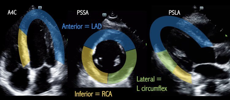
To evaluate for a regional wall motion abnormality (RWMA) remember the acronym SALPI (image 1). In the parasternal short axis view, starting at the septum, go clockwise to identify the anteriorlateralposteriorinferior walls. To look for a RWMA, look closely at each wall during systole to see if:

  1. The myocardium is moving in towards the center of the ventricle

  2. The myocardium is increasing in thickness

The absence of these findings is concerning for a RWMA which may be indicative of an acute MI. Patients with old MIs may also have RWMAs - correlate with the EKG and old echos if available

The parasternal long axis view and apical 4 chamber views can also be used to evaluated for RWMA (image 2).

When to POCUS for RWMA

This may be especially helpful in patients w/ equivocal EKGs that you or cardiology is on the fence about activating the cath lab or when the symptoms are not quite consistent with an MI (as in this case). Finding a RWMA may expedite cath lab activation (1).

Pro Tip: Cover up the entire LV with your hand except the specific wall you’re looking at and look at each wall seperately.

Back to the patient

The patient did not have any active chest pain but the initial troponin returned at 27.

The patient was taken to the cath lab which showed triple vessel disease with 80% stenosis of mid-LAD, 95% stenosis of first diagonal, 95% stenosis of proximal circumflex. He was evaluated for CABG but ultimately chose medical therapy.

References:

(1) Xu C, Melendez A, Nguyen T, Ellenberg J, Anand A, Delgado J, et al. Point-of-care ultrasound may expedite diagnosis and revascularization of occult occlusive myocardial infarction. Am J Emerg Med. 2022;58:186–91.


Joint Aspiration: Ankle

When to tap?

When you have a debilitating ankle injury with swelling at the tibiotalar joint preventing range of motion at that joint ie: dorsiflexion/plantarflexion.

What about the differential?

Ankle arthrocentesis allows for rapid identification of septic arthritis vs. gout vs. pseudogout vs. osteoarthritis vs. rheumatoid arthritis.

What are your landmarks?

The goal is to avoid the Dorsalis pedal artery, the peroneal nerve and the tendon of the Extensor Hallucis Longus (EHL). It is recommended to use an anterolateral approach where the joint line can be found between the lateral edge of the EDL and the medial edge of the lateral malleolus (Yellow Arrow Image 1). Plantarflex the ankle while the patient is bent at the knee in the supine position to widen the joint space prior to performing the procedure.

IMAGE 1:

Foot.jpg


How do you perform it?

  • 1. Patient should be in a supine position with the ankle in plantar flexion with plantar surface flat on the bed.  

  • 2. Mark you landmarks (see above).

  • 3. Prepare the site (ex. chloraprep)

  • 4. Anesthetize the area with smaller needle(23/25 gauge) creating a wheal and then advance creating the start of a projected path towards the joint capsule.

  • 5. Attach a 5 or 10 cc syringe to a 20 or 22 gauge needle and advance the needle into the joint space pulling negative pressure as you advance. The needle should be directed perpendicular to the tibia. If your syringe starts to fill up, and you need to get more fluid out, change out your syringe using hemostats to hold the needle. Most wrist and ankle effusions will yield only 1-3cc of fluid.

What about Ultrasound Guidance?

YES. This can absolutely be used to assist you in performing the procedure and will allow for visualization of your needle tip during aspiration.


For ultrasound guidance an anteromedial approach is generally used.

Landmarks- Place probe in between the TA tendon and EHL tendon, then rotate longitudinally with the probe marker facing the patient’s head  (Blue Arrow IMAGE 1). You will actually be inserting your needle medial to the TA tendon (Red Arrow IMAGE 1).

Image 2: 

more-tips-and-tricks-6.jpg
more-tips-and-tricks-7.jpg

Image 3:

AnkleTap.png