POTD: Blunt Cerebrovascular Injury

Todays POTD will be a trauma topic we frequently talked about at Shock Trauma but less frequently at Maimo.

 Blunt Cerebrovascular injury (BCVI): refers to a spectrum of injuries to the cervical carotid and vertebral arteries secondary to blunt trauma.

 Why is this Important?

If left untreated, patients with BCVI are at increased risk for stroke. Mortality may reach as high as 43%. Rare diagnosis which makes it even more important to consider evaluating for in all of your trauma patients that meet criteria and have the associated risk factors .

 Pathology:

The injury is caused by longitudinal stretching and injury to the vessels. Acceleration and deceleration can cause rotation and hyperextension of the neck, stressing the craniocervical vessels. This will lead to disruption of the intima. The intima tear then becomes a source of platelet aggregation that has a potential to cause downstream effects such as an embolic stroke or vessel occlusion

 Risk Factors:

·      High energy transfer mechanisms

·      LeFort II or III fractures

·      Mandibular fractures

·      Complex skull fracture/basilar skull fracture/occipital condyle fracture (most common risk facture)

·      Closed head injury with GCS < 6

·      Cervical spine fracture, subluxation, or ligamentous injury at any level

·      Near hanging with anoxic brain injury

·      Clothesline type injury or seat belt abrasion with significant swelling, pain, or AMS

·      Traumatic brain injury with thoracic injuries

·      Scalp degloving

·      Blunt cardiac rupture

·      Upper rib fractures

 Signs/Symptoms:

·      Arterial hemorrhage from neck/nose/mouth

·      Cervical bruit in patient < 50 years old

·      Expanding cervical hematoma

·      Focal neurologic defect

·      Neurologic defect inconsistent with CT head findings

·      Stroke on CT or MRI

 Diagnostics:

  • ·      Standard of care CTA (80% sensitive and 97% specific)

  • Should be considered when patient has one or more of the risk factors or signs and symptoms

  • ·      Can also do MRI or arteriography but this is time consuming and labor intensive

 Grading Scale:

1.     Grade 1: Intimal irregularity or dissection < 25 % of luminal narrowing noted

2.     Grade 2: Dissection or intraluminal hematoma with > 25% luminal narrowing, intraluminal clot or visible intimal flap

3.     Grade 3: Pseudoaneurysm

4.     Grade 4: Complete occlusion

5.     Grade 5: Transection with active extravasation

 Management:

·       Antithrombotics (heparin) or Antiplatelets (aspirin, Plavix) 

·       Operative repair

·       Endovascular stenting

·       Grade 1 and 2 injuries: single antiplatlet agent (aspirin 81 or 325mg)

·       Grade 3: dual antiplatelets or therapeutic anticoagulation (heparin drip with PTT at goal)

·       Grade 4 and above: Dual antiplatelets or therapeutic anticoagulation as well as operative or endovascular intervention

·       Many low grade injuries heal within 7-10 days therefore early repeat CTA is recommended. Otherwise treatment may need to be continued for 3-6 months.

 References:

·      https://www.emra.org/emresident/article/blunt-cerebrovascular-injury/

·      https://rebelem.com/blunt-cerebrovascular-injury-bcvi-universal-imaging-for-all/

·      https://jss.amegroups.com/article/view/3790/html

·      https://radiopaedia.org/articles/blunt-cerebrovascular-injury?lang=us

·      https://www.east.org/education-career-development/practice-management-guidelines/details/blunt-cerebrovascular-injury

·      https://www.aliem.com/guideline-review-east-blunt-cerebrovascular-injury/


 · 

POTD: Testicular Dislocation

Today’s Trauma Tuesday POTD is about a rare but dangerous type of straddle injury in males: testicular dislocation.

Most commonly found in young males involved in a decelerating motorcycle accident, testicular dislocation presents with severe unilateral or bilateral scrotal and/or inguinal pain. Because there may be multiple other distracting injuries incurred as a result of the accident, a genitourinary exam will be vital in identifying this injury.

On exam, you will find that the testicle has been dislocated from its normal home in the scrotum to another location due to blunt force tearing the fascia of the spermatic cord. Half of the time, that location is the inguinal pouch, and you may find a palpable mass representing the testis at the inguinal crease. The corresponding hemiscrotum will be empty. Interestingly, unilateral and bilateral testicular dislocation appears to occur at the same rate, so don’t forget to check the other side as well. Other locations the dislocated testis may end up are the penis, the perineum, and the abdomen.

Manual reduction may be attempted but is often limited by intractable pain and therefore infrequently successful. There also may be concomitant torsion. Emergent urology consult for operative intervention is usually indicated.

Prolonged dislocation may affect fertility and increases the risk of testicular malignancy in the future


Sources:

Zavras N, Siatelis A, Misiakos E, Bagias G, Papachristos V, Machairas A. Testicular Dislocation After Scrotal Trauma: A Case Report and Brief Literature Review. (2014) Urology case reports. 2 (3): 101-4.

S. L. Schwartz, G. Faerber. Dislocation of the testis as a delayed presentation of scrotal trauma. (1994) Urology.


 · 

POTD: Eye Stuff (Trauma Tuesday!)

POTD: Eye Stuff (Trauma Tuesday!)

A.

Seidel's sign: Fluorescein stained vitreous flowing from the site of globe perforation. Should protect the globe from any external pressure with eye shield, elevate head of bed 30 degrees, analgesia, control hypertension, and prevent vomiting. Emergent Optho consult.

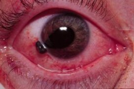
B.

Teardrop pupil: Usually indicated globe rupture/ FB. See management for Seidel's sign above.

C.

Corneal foreign body with rust ring: remove foreign body, urgent follow up for rust ring removal which should be done after 24 hours from initial injury- this is because reepithelialization makes removal easier.

D.

Exophthalmos: if in setting of trauma with increased intraocular pressure suspect retrobulbar hematoma. Obtain STAT CT scan, perform STAT lateral canthotomy and emergent optho consult.

E.

Hyphema: Blood in the anterior chamber of the eye. Elevate head of bed, control intraocular pressure. Patients on anticoagulation or antiplatelet agents should be admitted for reversal and observation. Consult ophthalmology depending on size of hyphema and rebleed risk.

Stay well,

TR Adam

A.

A.

B.

B.

C.

C.

D.

D.

E.

E.

 ·