POTD: Tongue Blade Test in Minor Mandibular Blunt Trauma

Minor trauma with mild swelling and want to avoid imaging the patient?

Tongue blade test:

How is it done? Have the patient attempt to "clamp down on” a tongue blade between the teeth with enough force that the examiner is unable to pull it out from the teeth.

When the examiner twists the blade, a patient should be able to generate enough force to break or crack the blade.

A positive test: if the patient cannot clench the tongue blade between the teeth or if the examiner cannot break the blade while it is held in the patient’s bite. If the test is positive, imaging is indicated.  

A negative test: If the blade can be gripped by the patient and be broken by the examiner, fracture of the mandible is much less likely, and additional imaging is likely not needed. In a prospective series of 110 patients with suspected mandible fracture, the test was found to be approximately 96% sensitive and 65% specific.

Who is not likely to benefit from this test? Major trauma that would indicate further imaging, signs of mandibular fracture such as: intraoral bleeding, tooth malocclusion, trismus, ecchymosis, and intraoral swelling.

Sources: https://www.aliem.com/2010/07/trick-of-trade-tongue-blade-is-as/

^ Check out this awesome aliem post and especially for the video demonstration

Alonso L, Purcell T. Accuracy of the tongue blade test in patients with suspected mandibular fracture. J Emerg Med. 1995;13(3):297-304. [PubMed]

Peer IX

 · 

POTD: Trauma Tuesday: Lateral Canthotomy

When to perform it?

·      To release orbital compression syndrome, most commonly due to retrobulbar hematoma.

·      IOP > 40, the pressure that indicated that you need to cut and release the compartment syndrome.

·      Without decompression, irreversible vision loss due to increasing orbital pressure may occur in as little as 90-120 minutes.

Clinical situation: trauma to the head/face.

Physical exam:

https://www.emra.org/emresident/article/emergency-department-evaluation-of-blunt-orbital-trauma/

https://www.emra.org/emresident/article/emergency-department-evaluation-of-blunt-orbital-trauma/

CT head and face

112-208-f2.jpg

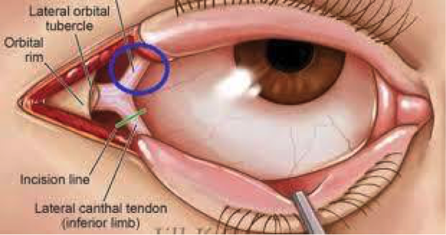
familiarize yourself with the anatomy

potd anatomy eye.png

Before starting, highly consider sedation.

Steps:

1)    Generously inject 1% Lidocaine with epi to numb that lateral canthus to the orbital rim.

  • Do this by directing the needle away from the globe itself

  • Helps with bleeding and with pain.

2)    Using the needle driver as your hemostat, advance from the lateral canthus to outer orbit rim. Clamp down and hold for 1- 2 minutes.

3)    Using your small scissors, cut the lateral canthus to the orbital rim.

4)    Then cut inferiorly to cut the inferior crus of the lateral canthus (you may need to probe around to feel the structures)

5)    Repeat IOP. If the IOP is not immediately lower, then cut the superior crus of the lateral canthus and recheck pressure.

Pearls:

·      There is a lot of swelling. It can be hard to fit your hemostat in place and to feel your landmarks. Use you instruments to feel/probe around.

·      Also, do not worry about cutting too much. You are doing this to save this patient’s vision. After discussing this with optho, these are repaired quite easily days/weeks later. For example, Dr. Tome Levy performed this once on a patient that I later followed up with in split flow about 2 weeks later. There was no physical evidence on physical exam that this had ever happened. At first I thought I had the wrong patient in front of me… but the patient confirmed that a week and half ago he had in fact had the optho plastic surgery to repair it.  

Sources:

https://emedicine.medscape.com/article/82812-overview

http://www.tamingthesru.com/blog/annals-of-b-pod/ocular-emergency

This is an excellent emrap video that reviews the procedure: https://www.youtube.com/watch?v=tgQaKVGynFA

 · 

POTD Trauma Tuesday: Name that fracture!

A 36-year-old man presents by ambulance following a motorcycle crash. He told the EMTs that he lost control and fell sideways, bracing his fall with his outstretched right hand. His R arm looks deformed but is neurovascularly intact. An xray is obtained.

xray potd.png


What’s the name of this one again?!

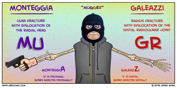
Galeazzi fracture! Named after an Italian surgeon from Milan.

What is it? a fracture-dislocation of the distal third of the radius associated with dislocation-subluxation of the distal radial ulnar joint, or DRUJ.

Why do we care? With Galeazzi fractures, there is a high risk of malunion, loss of function, infection, and chronic pain in adult patients. For this reason, surgical management with internal fixation and possible open reduction is required. The repair should occur promptly, so the emergency physician or another clinician should contact the orthopedic consultant emergently to coordinate care.

What about in peds? Emergent orthopedic consultation is still required but interestingly, in children, some Galeazzi fractures are treated conservatively with closed reduction by an orthopedic surgeon. Disruption of the DRUJ can be subtle, so a high suspicion should be maintained when a patient presents with a fracture of the distal third of the radius.

Pearls of the Peal:

* Look for fracture-dislocation of the distal radius and ulna after a fall onto an outstretched arm. This injury can’t be missed: it requires immediate orthopedic involvement.

* Skin tenting associated with the Galeazzi fracture-dislocation puts the patient at risk for skin necrosis and conversion to an open fracture.

Wasn’t there some way to remember this compared to other one?? Why, yes! See below:


mugger.png

Sources:

Comic: Medcomic.com

Xray and clinical information: PEER IX

 ·