COPD and antibiotics.

Welcome back to POTD. 

The weekend has come and the weekend has gone. I know you've all been holding your breath to hear about----

A message from our sponsors:

Take a deep inhale. feel some wellness. feel the firmness of your feet on the floor. hold onto your seat.   

Exhale nice and slowly......like someone with a COPD exacerbation.

Because today we're discussing antibiotic coverage in acute COPD Exacerbations. I know you've been waiting a lung time for this one. 

Background

  • Acute COPD exacerbations (AECOPD) account for ~1.5 million ED visits annually in the ED.

  • Many physicians routinely prescribe antibiotic coverage for AECOPD

  • a 2018 review demonstrated antibiotic prescriptions given on 39% of ED visits for AECOPD between 2009-2014.

  • Due to the structural changes in the bronchi of COPD patients they are more prone to bacterial colonization (as opposed to asthmatics - which have no structural change but a reactive process)

Do guidelines exist?

  • Sure do. 

  • if the patient appears infectious (think fever) administer antibiotics. This is understandable given their risk factors and bronchial structural changes.

  • Several guidelines exist for more subtle cases, they exist as follows: (see chart below)

    • Global initiative for Chronic Obstructive Lung Disease:

      • Antibiotics should be given to

        • patients with all 3 of the following cardinal symptom

          1. increased dyspnea

          2. increased sputum volume

          3. increased sputum purulence

        • patients with 2 cardinal symptoms, if there is increased purulence

        • patients requiring noninvasive or invasive ventilation

    • American Thoracic Society/European Respiratory Society

      • hospitalized patients with chanegs in sputum characteristics

      • all patients admitted to an ICU

    • Canadian Thoracic Society

      • patients with severe purulent AECOPD

    • National Institute for Health and Clinical Excellence

      • patients with more purulent sputum

  • Basically, pay attention to that sputum. take a thorough history and discuss changes in sputum production. 

-Elly

 · 

ARDS

Screen Shot 2019-08-20 at 8.55.51 PM.png

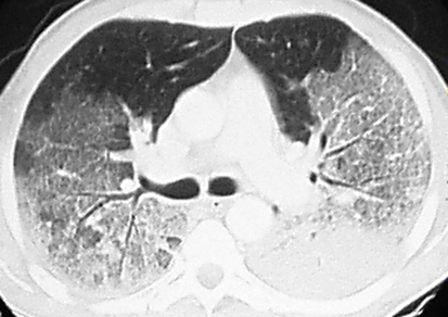
Acute respiratory distress syndrome (ARDS) 

acute inflammatory lung injury that causes non-cardiogenic pulmonary edema by increasing alveolar capillary permeability. 

The thickened diffusion barrier leads to hypoxemia via:

decreased lung compliance

inefficient gas exchange

Pulmonary hypertension

increased physiological dead space

Predisposing factors:

Direct lung injury: pneumonia, gastric aspiration, pulmonary contusion, near drowning, inhalation injury, transfusion-related acute lung injury

Indirect lung injury: sepsis, shock, acute pancreatitis, burns, crush injury, fat embolism, and massive transfusion

Diagnosis criteria for ARDS – Berlin definition (all 4 components must be present):

  1. Acute onset (1 week or less)

  2. Hypoxemia (PF ratio* < 200 mmHg with a minimum of 5 cmH2O PEEP (or CPAP))

  3. Pulmonary edema (bilateral opacities on CXR)

  4. Non-cardiogenic (not caused by cardiac failure)

*PF (PaO2/FiO2) ratio is the ratio of arterial oxygen partial pressure to fractional inspired oxygen. PaO2 value can be obtained from ABG, and FiO2 is 0.21 at sea level (room air) or depends on supplemental O2.


ARDS is a diagnosis of exclusion so consider first: 

Cardiogenic pulmonary edema, severe multilobar pneumonia, acute exacerbation of pulmonary fibrosis, diffuse alveolar hemorrhage, idiopathic acute eosinophilic pneumonia, dissemination of lymphoma/leukemia, and several others. 


Workup:

Labs: CBC, BMP, LFTs, Coags, VBG followed by ABG, troponin, BNP, lipase, consider DD

Imaging: CXR, POCUS US ECHO and CHEST and consider CT


ED Management:

Supplemental O2

Treat the underlying condition (pneumonia, sepsis, etc.)

Tempered diuresis – non-cardiogenic pulmonary edema takes much longer to respond to treatment than cardiogenic CHF, so avoid being overly aggressive with diuresis, as this may worsen underlying shock and increase likelihood of multi-organ failure

Glucocorticoids — consider steroids when ARDS precipitated by a steroid-responsive process (eg, acute eosinophilic pneumonia)

Be cautious when using non-invasive positive pressure ventilation – the benefit of NIPPV in the initial management of ARDS remains controversial. 

Mostlikely patient will end up being intubated, for vent management suggested strategies are:

Use low tidal volume (6-8 mL/kg) to avoid barotrauma (ideal body weight should be calculated)

And careful FiO2:PEEP ratio titration:


Screen Shot 2019-08-20 at 11.28.45 AM.png


ARDS severity (mortality) predictor 

Mild ARDS – The PaO2/FiO2 is >200 mmHg, but ≤300 mmHg, on ventilator settings that include positive end-expiratory pressure (PEEP) or continuous positive airway pressure (CPAP) ≥5 cm H2O

ARDS_CT.jpeg

Moderate ARDS – The PaO2/FiO2 is >100 mmHg, but ≤200 mmHg, on ventilator settings that include PEEP ≥5 cm H2O

Severe ARDS – The PaO2/FiO2 is ≤100 mmHg on ventilator settings that include PEEP ≥5 cm H2O.


systematic approach to reading CXR and hidden pneumonias


For this pearl of the day we will talk about systematic approach to reading CXR and hidden pneumonias:


The key is to be very systematic when approaching CXRs and that is what radiologists do each time.

Here is the suggested approach by the Brown EM program (https://brownemblog.com/?offset=1533674064239&category=Education)

Screen Shot 2019-08-16 at 2.07.54 PM.png


https://commons.wikimedia.org/wiki/File:Mediastinal_structures_on_chest_X-ray.svg#/media/File:Mediastinal_structures_on_chest_X-ray,_annotated.jpg

When ready to review the x-ray, consider the commonly used “A, B, C, D, E, F” system.

A - Airway- trachea, carina, right and left main bronchi

B - Bones and soft tissue- clavicles, ribs- posterior and anterior, vertebral bodies, and sternum on lateral films. Look for any fractures, dislocations, or lytic lesions.

C - Cardiac- cardiac silhouette and mediastinum. The cardiac silhouette should be less than half of the thoracic cavity. AP films exaggerate heart size, so this rule does not apply. Assess the borders of the heart and the hilar structures

D - Diaphragm- right should be higher than left and you should see a gastric air bubble on the left. Is there any free air under the diaphragm? Evaluate the costophrenic angle and pleura (normally invisible due to thinness).

E - Everything else (lines and tubes, pacemakers, artificial valves)

F - Fields- FINALLY, evaluate the lung fields. Lungs are the area of greatest interest, so it is helpful to keep this at the end to prevent distraction. Divide each lung into three “zones” when reading a chest x-ray. These do not correlate with the lobes. Remember, there are 2 lobes on the left (upper and lower) and 3 on the right (upper, middle and lower). 


Hidden pneumonias:

Go through your ABCDEFs and look at the signs of hidden pneumonias:


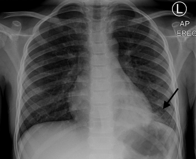
Silhouette sign

The loss of the normal silhouette of a structure is called the silhouette sign.  - It enables us to find subtle pathology and to locate it within the chest.

Screen Shot 2019-08-16 at 2.09.18 PM.png


Screen Shot 2019-08-15 at 4.16.26 PM.png

R middle lobe pneumonia

2a.jpeg

LLL pneumonia

Screen Shot 2019-08-15 at 4.27.05 PM.png

LLL pneumonia


Hidden areas

There are some areas that need special attention, because pathology in these areas can easily be overlooked:

apical zones

hilar zones

retrocardial zone

zone below the dome of diaphragm

These areas are also known as the hidden areas.


But in doubt get another view or a chest CT.


References:

https://brownemblog.com/?offset=1533674064239&category=Education

http://www.radiologyassistant.nl/en/p497b2a265d96d/chest-x-ray-basic-interpretation.html#in5145a34e91e18

https://www.bir.org.uk/media/258608/mark_rodriguez_-_philips_trainee_for_excellence_-_unofficial_guide_to_radiology.pdf