VOTW: HAND me the probe and let me FLEX my POCUS skills!

HPI: 44 year old male with no PMH presenting to the ED for worsening left 3rd finger pain and swelling after sustaining trauma and laceration to affected area 9 days ago. The team's differential included finger cellulitis, abscess, flexor tenosynovitis, and underlying fracture.

The patient’s hand was placed in a water bath and the following images were obtained using the linear probe:

POCUS evaluation of flexor tenosynovitis

  1. Use a water barrier between probe and fingers to improve image quality(ex: plastic basin, emesis bag, glove filled with water, bag of NS/LR).

  2. Use the linear probe on the flexor side of the fingers.

  3. Evaluate the flexor tendon which overlies the bone. Look for fluid (anechoic) within the flexor tendon sheath surrounding the flexor tendon. Remember, tendons are anisotropic which means they can appear hyperechoic or hypoechoic depending on the angle of your probe. Hypoechoic areas can be confused for edema so it is important to fan through the entire tendon. If the area of concern remains consistently hypoechoic, that is more concerning for fluid/edema.

  4. The tendon may also appear thicker compared to fingers. If you apply color doppler, you may see surrounding hyperemia.

  5. You can scan an unaffected finger also for real time comparison on what “normal” should look like.

Case conclusion: After this bedside POCUS, orthopedics team was consulted for concern for flexor tenosynovitis!

Learn more about POCUS findings for flexor tenosynovitis here:

  1. https://coreultrasound.com/fts/

  2. https://www.ultrasoundgel.org/posts/q08ayJgg3rmHtiQgs9n82w


VOTW: Mass-ive Fever

This weeks’ VOTW was brought to you by Drs. Hannah Blakely, Patricia Camino, and the ultrasound team that was scanning that day!

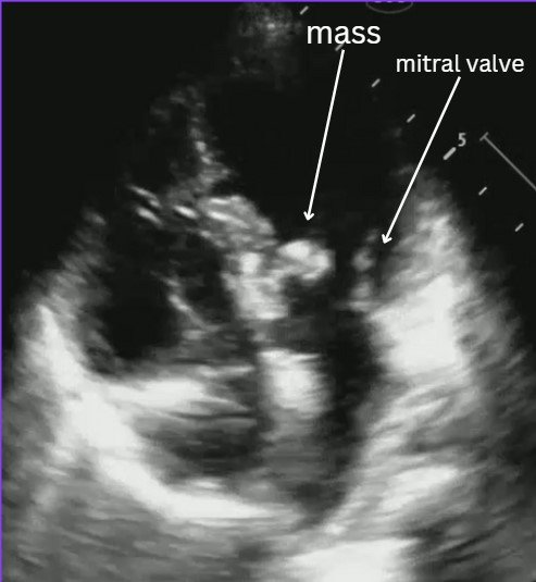
HPI: 6 year old female with PMH of atrioventricular canal defect s/p repair, recent strep throat infection presenting for fever x 14 days.

Bedside POCUS showed:

Evaluating for endocarditis on POCUS:

B mode: look for masses, usually on the lower flow side of the valves (ex: mitral valve- endocarditis is more likely to be found on atrial side).

Color flow: you can usually find associated regurgitation of the affected valve

Possible mimics:

  • Thrombus

  • Papillary muscle rupture/flail leaflet

  • Intracardiac tumor

  • Artifact

Remember that a valve vegetation is one of the major diagnostic criteria for endocarditis. In the right clinical scenario this POCUS finding can highly increase your suspicion for endocarditis.

Case conclusion: CTAP significant for possible splenic and renal infarcts. Patient was admitted for suspected endocarditis. Blood cultures were +MSSA. Pediatric cardiology ECHO was consistent with mitral valve vegetation consistent with endocarditis and septic emboli.

Resources for more info:


Happy scanning!

  • The US Team


VOTW: You take my breath away!

HPI: 90 yo female presenting for worsening shortness of breath and tachycardia x 3 days and right leg pain x 2 weeks with difficulty ambulating.

POCUS showed:

ECHO A4C view (see video): note the size of the RV appears larger than the LV. This is a sign of right heart strain and in the appropriate setting (such as this one) can be concerning for a pulmonary embolism!

Compression views of the common femoral vein (CFV), femoral vein (FV), and popliteal vein (PV). See the echogenic material inside the popliteal vein which is suggestive of a DVT. Remember that during the acute phase of a DVT (<14 days), the clot may appear isoechoic to the blood inside the vein so you may not see this echogenic material and should rely more on your compression exam.

Review on how to do DVT US:

Linear probe

Patient in frog leg position

4 main areas to view

  • Common femoral vein (CFV)-saphenous vein junction (SFV)

    • Clot noted in the SFV within 3 cm to the junction is treated as a DVT. More distally, if there is 5 cm worth of clot noted in the SFV it is also treated as a DVT.

  • CFV branching into [superficial] femoral vein and deep femoral vein

  • Mid/distal femoral vein

  • Popliteal vein

    • Remember the popliteal vein is on top of the popliteal artery (pop on top!)


Tips:

  • You often have to go much higher in the groin than you think to find the CFV-SFV junction

  • Compression testing of the deep veins should not compress the artery (if it is, you’re pressing too hard and can miss subtle DVTs)

  • Deep veins are paired with arteries so identify your landmarks to ensure you are looking at the correct vessels

  • Use your non-scanning hand to help with compression of deeper veins by supporting the other side of the patient’s leg

Case conclusion: Patient with elevated troponin and BNP. CTA significant for bilateral PE’s. Labs and ECHO findings consistent with submassive PE. Patient started on heparin drip and admitted to the floor!

Happy scanning!

  • The US Team