POTD Trauma Tuesday: Name that fracture!

A 36-year-old man presents by ambulance following a motorcycle crash. He told the EMTs that he lost control and fell sideways, bracing his fall with his outstretched right hand. His R arm looks deformed but is neurovascularly intact. An xray is obtained.

xray potd.png


What’s the name of this one again?!

Galeazzi fracture! Named after an Italian surgeon from Milan.

What is it? a fracture-dislocation of the distal third of the radius associated with dislocation-subluxation of the distal radial ulnar joint, or DRUJ.

Why do we care? With Galeazzi fractures, there is a high risk of malunion, loss of function, infection, and chronic pain in adult patients. For this reason, surgical management with internal fixation and possible open reduction is required. The repair should occur promptly, so the emergency physician or another clinician should contact the orthopedic consultant emergently to coordinate care.

What about in peds? Emergent orthopedic consultation is still required but interestingly, in children, some Galeazzi fractures are treated conservatively with closed reduction by an orthopedic surgeon. Disruption of the DRUJ can be subtle, so a high suspicion should be maintained when a patient presents with a fracture of the distal third of the radius.

Pearls of the Peal:

* Look for fracture-dislocation of the distal radius and ulna after a fall onto an outstretched arm. This injury can’t be missed: it requires immediate orthopedic involvement.

* Skin tenting associated with the Galeazzi fracture-dislocation puts the patient at risk for skin necrosis and conversion to an open fracture.

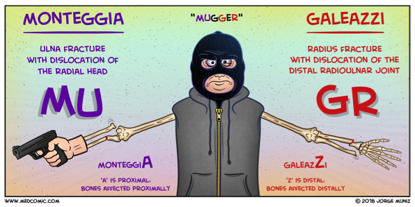
Wasn’t there some way to remember this compared to other one?? Why, yes! See below:


mugger.png

Sources:

Comic: Medcomic.com

Xray and clinical information: PEER IX

 · 

POTD: Straight leg test. A leg up on clinical testing!

A little background:

Lumbar disc herniation is the most common cause of lumbar radiculopathy, or sciatica, a shooting or burning pain from the low back radiating down the posterior leg distal to the knee.

Two tests used to evaluate these symptoms are

The straight leg raise.

·       The straight leg raise test is highly sensitive but not very specific for disc herniation.

·       This is performed by lifting the leg affected by the radiating pain.

·       The patient lies supine with one leg either straight or flexed at the knee with the sole of the foot flat on the stretcher.

·       The examiner then raises the affected leg up, extended, to 30 to 70 degrees.

·       Reproduction of low back pain that radiates down the posterior affected leg at least past the knee is considered a positive result. Not just pain to the lower back, which is a common misconception.

·       The SLR test can also be performed with the patient in a sitting position, by stretching the sciatic nerve by extending the knee; the test is positive if pain radiates to below the knee.


The crossed straight leg raise.

·       It is highly specific (90%) for disc herniation

·       You perform the same test as the straight leg but on the unaffected leg.

·       A positive test: reproducing both the back pain + the radiation down the affected leg.

Sources: Peer IX, Tintinelli’s, Dr. Sergey Motov, Uptodate

 · 

Hip Dislocations POD

This POD was inspired by a case that Dr. Zerzan had in the Peds ED. An 8 year old with a traumatic injury presented with hip pain and was found to have an isolated posterior hip dislocation…

PastedGraphic-7.tiff
PastedGraphic-6.tiff
PastedGraphic-5.tiff
PastedGraphic-4.tiff
PastedGraphic-3-1.tiff
PastedGraphic-2-1.tiff

Hip dislocations!

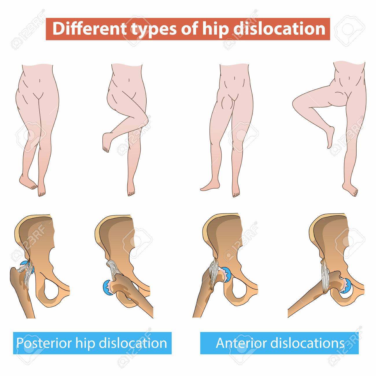
Posterior hip dislocations (PHDs) are far more common than anterior hip dislocations

(90% - 10%). This holds true in pediatrics as well in adults.

PastedGraphic-1.jpeg

In a posterior dislocation, the patient presents with the extremity internally rotated and shortened.

PastedGraphic-2.jpeg
PastedGraphic-4.jpeg

In anterior dislocations, patients typically present with extremity flexed, abducted, and externally rotated.

PastedGraphic-3.jpeg
PastedGraphic-5.jpeg

We will focus on

posterior dislocations.

Classic presentation is with an axial load such as a knee hitting the dashboard in an MVC or other high energy mechanisms.

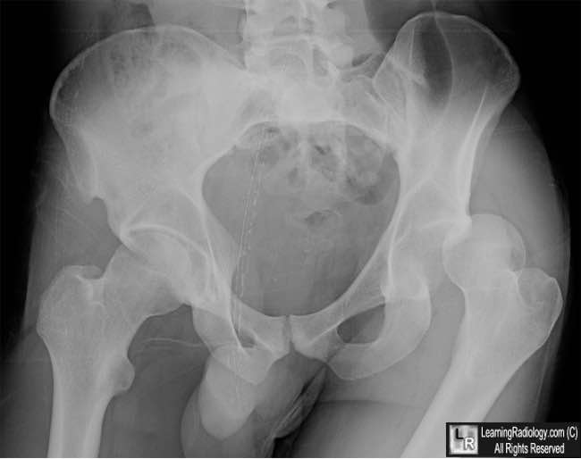
hip-dislocation-POD.jpg

Important point: in adults and children >10yo, PHDs require a high energy mechanism and will often have several associated injuries.

However in children <10yo, PHDs can be seen in lower energy mechanisms such as routine sports injuries which is why you may actually see an isolated hip dislocation in a child. There are also fewer associated acetabular fractures in pediatric PHDs than adult PHDs.

Any child PhD knows…

PastedGraphic-6.jpeg

..that PHDs are true emergencies!

You need to

get it reduced ASAP (within 6 hours)

to prevent complications of femoral head osteonecrosis and sciatic nerve injury. Other complications include post-traumatic arthritis, and in pediatrics, physeal injury. Incidence of recurrent dislocation is higher in pediatrics than in adults!

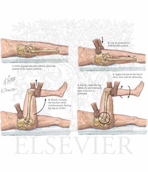
Reduction techniques:

The Allis Maneuver:

PastedGraphic-7.jpeg

The Captain Morgan:

PastedGraphic-8.png

Video here: 

https://www.youtube.com/watch?time_continue=82&v=lQMWaFX-MeQ

Propofol is preferred agent for procedural sedation given its muscle relaxant properties if it is going to be reduced in the ED, but pediatric cases are often reduced in the OR to ensure optimal muscle relaxation and to have more options available.

It is essential to have optimal muscle relaxation in pediatrics as the growth plates can be damaged during reduction.

Open reduction should be considered if fracture-dislocation or unsuccessful closed reduction attempt.

All patients should get at least a CT to evaluate for femoral head fractures, intra-articular loose bodies/incarcerated fragments, acetabular fractures.

Children should get an MRI to evaluate for ligamentous injury as well.

If closed reduction is successful, disposition is protected weight-bearing 4-6 weeks, ortho follow up.

PastedGraphic-1-1.tiff
PastedGraphic-2-1.tiff
PastedGraphic-3-1.tiff
PastedGraphic-4.tiff
PastedGraphic-5.tiff
PastedGraphic-6.tiff
PastedGraphic-7.tiff
 ·