UV keratitis

When you see someone chilling on a stretcher with sunglasses on in the middle of south this holiday weekend, they might not just be trying to sleep off the two dozen margaritas they just had.

What is UV keratitis? It’s bilateral eye pain usually 30 min to 12 hours after UV exposure, think of it as a “sunburn” of the eyes. Your patient was lounging on the beach all day Saturday and used a pair of knock-off sunglasses or those free ones they hand out at career fairs. This can also happen in the winter for skiers on a bright sunny day and all that sunlight reflecting off the snow into their eyes. Other sources: tanning beds, arc welding, laboratory UV lights

Pathophysiology: cornea absorbs UV light à epithelial cell death/desquamation à symptoms resolve when corneal epithelium regenerates

Symptoms: conjunctival injection, photophobia, foreign body sensation, inability to open eyes, facial erythema/edema

Exam:

normal or reduced visual acuity

bulbar conjunctival (the covering of the white part of the eye) injection and chemosis with sparing of the palpebral conjunctiva since it is blocked by the eyelids

punctate keratitis on Fluorescein stain!

UV keratitis fluorescein stain.jpg

Rx:

Supportive care, similar to corneal abrasion management

Tell the patient that healing should occur within 24-72 hours

Topical antibiotic ointments => Consider Erythromycin or Polymixin-Bacitracin 3-4 times daily for 2-3 days

Stay away from topical anesthetics, no they cannot take home that bottle of tetracaine you used to numb their eye for the fluorescein exam => Risk of neurotrophic ulceration due to lack of protective reflexes (tearing and blinking)

Refer to Ophthalmologist if persistent signs and symptoms > 72 hours

Sources

https://eyewiki.aao.org/Photokeratitis

https://wikem.org/wiki/Ultraviolet_keratitis

 · 

POTD: Idiopathic Intracranial Hypertension

POTD: Idiopathic intracranial hypertension


Idiopathic intracranial hypertension (IIH) aka pseudotumor cerebri and benign intracranial hypertension

·      rare condition

·      presents with gradual onset and chronic headache, vision changes, nausea, vomiting, and tinnitus

·      + papilledema/ swelling of the optic disc on fundoscopy

potd eye papill.jpg

·      optic sonography

potd us eye.jpg
  • ONSDs should be measured 3 mm behind the papilla, an average of less than 5 mm is considered normal.

  • ONSD > 5 mm has been shown to be 90% sensitive and 85% specific for ICP > 20.

·      Classic presentation: young, obese female

·      + association has been found with this diagnosis and the use of oral contraceptive medications, tetracycline, anabolic steroids, and vitamin A

·      Pathophysiology is not well understood but thought to be caused by an imbalance in CSF production and reabsorption

·      Diagnostic criteria include an alert patient with either a normal neurologic examination or findings consistent with papilledema, visual field defect, or an enlarged blind spot

·      Definitive dx: Lumbar puncture

  • done in a recumbent position reveals an elevated CSF opening pressure of more than 20 mm Hg in an obese patient (normal being up less than 20 mm Hg).

  • normal CSF analysis.

·      CT head may show “slit like” or normal ventricles without mass effect

·      DDx: glaucoma, venous sinus thrombosis, ICH, IC mass.

·      Treatment

  • Repeat LPs  

  • Acetazolamide

  • Surgical shunt if severe and refractory

  • offending agents such as oral contraceptive medications should be discontinued.

·      Permanent loss of vision can occur in up to 10% of patients, and higher if left untreated


Sources:


  • Dubourg J, Javouhey E, Geeraerts T, Messerer M, Kassai B. Ultrasonography of optic nerve sheath diameter for detection of raised intracranial pressure: a systematic review and meta-analysis. Intensive Care Med. 2011;37(7):1059-68. [pubmed]

  • Blaivas M, Theodoro D, Sierzenski PR. Elevated intracranial pressure detected by bedside emergency ultrasonography of the optic nerve sheath. Acad Emerg Med. 2003;10(4):376-81. [PDF]

  • https://www.ultrasoundoftheweek.com/uotw-5-answer/

  • Peer IX

 · 

POTD: Trauma Tuesday: Lateral Canthotomy

When to perform it?

·      To release orbital compression syndrome, most commonly due to retrobulbar hematoma.

·      IOP > 40, the pressure that indicated that you need to cut and release the compartment syndrome.

·      Without decompression, irreversible vision loss due to increasing orbital pressure may occur in as little as 90-120 minutes.

Clinical situation: trauma to the head/face.

Physical exam:

https://www.emra.org/emresident/article/emergency-department-evaluation-of-blunt-orbital-trauma/

https://www.emra.org/emresident/article/emergency-department-evaluation-of-blunt-orbital-trauma/

CT head and face

112-208-f2.jpg

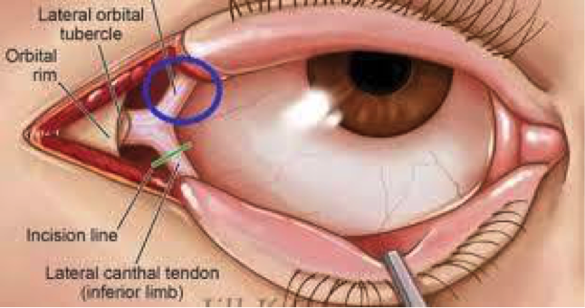
familiarize yourself with the anatomy

potd anatomy eye.png

Before starting, highly consider sedation.

Steps:

1)    Generously inject 1% Lidocaine with epi to numb that lateral canthus to the orbital rim.

  • Do this by directing the needle away from the globe itself

  • Helps with bleeding and with pain.

2)    Using the needle driver as your hemostat, advance from the lateral canthus to outer orbit rim. Clamp down and hold for 1- 2 minutes.

3)    Using your small scissors, cut the lateral canthus to the orbital rim.

4)    Then cut inferiorly to cut the inferior crus of the lateral canthus (you may need to probe around to feel the structures)

5)    Repeat IOP. If the IOP is not immediately lower, then cut the superior crus of the lateral canthus and recheck pressure.

Pearls:

·      There is a lot of swelling. It can be hard to fit your hemostat in place and to feel your landmarks. Use you instruments to feel/probe around.

·      Also, do not worry about cutting too much. You are doing this to save this patient’s vision. After discussing this with optho, these are repaired quite easily days/weeks later. For example, Dr. Tome Levy performed this once on a patient that I later followed up with in split flow about 2 weeks later. There was no physical evidence on physical exam that this had ever happened. At first I thought I had the wrong patient in front of me… but the patient confirmed that a week and half ago he had in fact had the optho plastic surgery to repair it.  

Sources:

https://emedicine.medscape.com/article/82812-overview

http://www.tamingthesru.com/blog/annals-of-b-pod/ocular-emergency

This is an excellent emrap video that reviews the procedure: https://www.youtube.com/watch?v=tgQaKVGynFA

 ·