POTD: Peripartum Cardiomyopathy

POTD: Peripartum Cardiomyopathy

Causes:

  • Infectious (EBV, CMV, HSV)

  • Genetics

  • Pre-eclampsia

  • Fetal cells present in the maternal system that elicit an inflammatory response

Clinical Findings (same as CHF findings):

  • Tachycardia

  • Decreased pulse oximetry (should be ≥ 97% at sea level).

  • Blood pressure may be normal. (systolic >140 mm Hg and/or diastolic >90 mm Hghyperreflexia with clonus suggest preeclampsia).

  • Elevated jugular venous pressure

  • Third heart sound (turbulent ventricular filling secondary to poor wall relaxation from dilated ventricle)

  • Loud pulmonic component of the second heart sound (increased right sided flow)

  • Mitral or tricuspid regurgitation

  • Pulmonary rales

  • Peripheral edema

  • Ascites

  • Hepatomegaly

Management:

  • CBC- to see if there is significant thrombocytopenia

  • CMP- to see if there is any dysfunction in creatinine, LFTs, albumin

  • Urine dipstick- to check if there is any proteinuria

  • EKG

  • Echocardiogram

  • CXR

  • Stress testing

  • OBGYN, Cardiology consult in addition to reaching out to potential transplant hospitals

Treatment:

  • Digoxin: first line in pregnancy

  • Loop diuretics; Start with 10 mg of furosemide, as pregnant women have an increased glomerular filtration rate (GFR) that facilitates secretion of the drug into the loop of Henle.

  • Hydralazine and nitrates: afterload and preload reduction

  • B- Blockers (carvedilol or metoprolol): decrease all-cause mortality and hospitalization in those with systolic dysfunction.

  • Heparin for EF<30% (high risk of venous and arterial thrombosis)

  • LVAD

  • May ultimately need heart transplant

  • Delivery- Unless the mother is decompensating, you can manage her medically until delivery is possible. If the mother is not responding to medical therapy or if the fetus must be delivered for obstetric reasons, the best plan is to induce labor with the goal of a vaginal delivery. C-section can lead to a lot of dynamic fluid changes which can lead to maternal decompensation

Disposition: 

  • ICU vs potential transfer to a center that offers tertiary care services for both the mother and the fetus.

Stay well,

TR Adam

 · 

EMS Protocol of the Week - Obstetric Complications

Short cooldown email for today’s EMS PotW (EMS-PoW? Is that catchier?). Protocol 540 – Obstetric Complications, attached below, shockingly addresses potential complications with the obstetric patient. For context, NYC REMAC is using a 160/110 cutoff for severe preeclampsia, associated with some sort of symptomatology. The protocol opens with reference to BLS procedures, which in this case is essentially just checking ABC’s, calling for ALS if needed, and considering IVC syndrome, moving the patient into a left lateral position if indicated. At this point, the only SO ALS intervention is IV access and fluids, and ultimately an OLMC call to you fine folks for discuss magnesium administration.

The MCO for magnesium is written as a 2 gram IV dose over 10-20 minutes, with a repeat dose of 2 grams if needed. Seeing as we normally start at 4 grams for our preeclampsia/eclampsia patients, this allows for an early start for the loading dose at a lower rate while getting the patient to the ED for further eval. Generally, I tend to have a low threshold for authorizing the magnesium, assuming the crew paints a picture of a patient who could benefit from it.

www.nycremsco.org and the protocol binder, both there for you when you need them most.

David Eng, MD

Assistant Medical Director, Emergency Medical Services

Attending Physician, Department of Emergency Medicine

Maimonides Medical Center

 · 

Intrauterine Devices

Studies show that about 10% of US women aged 15-49 use long acting reversible contraception (LARCs) and many of those happen to be intrauterine devices, or IUDs.

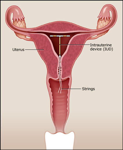
IUDs are a small flexible T-shaped piece of plastic that sit in the uterus and are used for contraception.

iudscehmatic.jpg

What do we as Emergency providers have to know about IUDs and when is it an emergency?

There are 2 categories of IUDs: copper and hormonal.

5 brands are approved for use in the US.

ParaGard (copper)

works by causing an inflammatory response which impairs movement and viability of sperm and diminishes the ability of a fertilized egg to implant

can be used for 10 years

women experience heavy periods/more cramping

string color is clear or white


Mirena, Liletta, Skyla, Kyleena (hormone)

works by releasing a progestin hormone which thickens the cervical mucus, thins uterine wall, and impairs binding of sperm to the egg

can be used for 3-5 years depending on the brand

women experience lighter periods/less cramps

string color is blue or brown


Complications are most commonly see within 1 month of placement and include PID, expulsion, perforation of uterus.


 How to evaluate a patient with an IUD coming in with lower abdominal/pelvic complaints:

 On exam, look for the IUD strings. There are 2 but they can appear as 1. They are generally 2.5cm in length.

 If the strings are too short or not visible at all:

The strings might be curled up in the cervix. Take a cytobrush or Q-tip and sweep the os to see if you can uncurl the strings.

If you still can't see the strings, perform a bedside ultrasound.

If the IUD is in the right location but there are no strings visible, provide outpatient OBGYN follow up.

goodiud.jpg

If the IUD is visible on ultrasound but not in the right location, it might have perforated the uterus. Call OBGYN to evaluate the patient.

If the IUD is not visible on ultrasound, it might have perforated entirely through the uterus. Perform a KUB to make sure it is not somewhere else in the abdomen. If you see it on KUB, again call OBGYN.

If you don't see it on KUB, it's possible that the IUD has fallen out of the uterus. Provide outpatient OBGYN follow up.

kubiud.jpg

If the strings are too long or absent and the IUD is partially out, remove the IUD completely.

If the patient has PID, treat it the same way you would if the patient did not have an IUD and provide OBGYN follow up. Although not removing an IUD in this case goes against logical sense, studies have shown that women who retained their IUDs had similar or better outcomes than women who had their IUDs removed.

Definitely still perform a pregnancy test. There is a 0.1-1.5% risk of pregnancy, depending on which brand of IUD. If the patient is pregnant, be very worried about an ectopic pregnancy and evaluate for it!

If the patient wants to keep her pregnancy and you see an IUP on ultrasound, keep the IUD in and provide OBGYN follow up, assuming patient is otherwise stable and ready to be discharged. Removing the IUD may actually disrupt the pregnancy.

If there is no IUP and patient is stable, asymptomatic, and being discharged, be sure to provide urgent OBGYN follow up.

To remove the IUD, you can use a pair of forceps to pull on the strings. There should be no resistance. If you meet any resistance, stop and call OBGYN.

When removing an IUD, remember to tell your patient that they can become fertile again within a couple days. If they recently had intercourse, sperm can be viable for 5 days, so offer them a Plan B pill.

 ·