Winter is coming, DE Winter

lets talk about two STEMI equivalents, this post covers De winters, Wellens, hypokalemia, hyperkalemia, to jump to a section look for the BOLDED headers

De Winter T waves

  • Indicates an acute proximal LAD occlusion

  • J point depression of 1-3mm

  • tall wide symmetric T waves

  • present in precordial leads v2-6

  • usually 0.5-1mm elevation aVR

de_wineter_st_t_graphic.jpg

Wellens syndrome

  • indicates acute or chronic proximal LAD stenosis

  • may present after chest pain

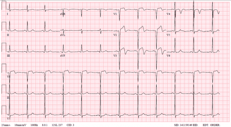
  • type A symetric isoelectric t waves in V1-2

  • type B has deep symetric T waves in V1-2

  • T changes maybe all the way to V6

  • typically without ST elevation

  • chest pain is often resolved

  • Do NOT stress test* patient has no collaterals patient requires catheterization!

type A:
type B:

wellens.png

*there are case reports of large anterior MI's after stress tests

Hypokalemia

  • Lengthened PR interval

  • T flattening/inversion

  • ST depression which can mimic ischemia

  • U waves after inverted T waves making the appearance of prolonged QT

  • Ectopic atrial and ventricular beats

  • Will progress to afib, vtach, vfib, or torsades de pointes

Hyperkalemia

  • prolonged PR

  • shortened QT

  • flattened P waves

  • Wide QRS

  • sharp symmetric peaked T waves

  • can have heart block ventricular arythmias

  • progresses to sinusoidal wave of WRS and peaked T and cardiac arrest

 · 

Not All ST Changes Are Created Equal

EMS rolls in performing CPR on your new patient an elderly man found down.  After achieving ROSC you receive the following EKG.  Where will you find this patient's lesion?

That is correct, this patient has an intracranial hemorrhage causing increased intracranial pressure.  Keep in mind up to 10% of cardiac arrests outside the hospital are from intracranial hemorrhage.   Typical ECG characteristics are:

  • Widespread deep wide T waves (cerebral T waves)

  • Qtc prolongation

  • Bradycardia

less common findings include:

  • St depression/elevation

  • Increased U wave amplitude

  • Other rhythm derangements such as premature ventricular contractions or afib

Keep in mind ICH can cause wall motion abnormalities visible on echocardiogram

The following ecg with diffuse ST elevations is from a trauma patient with ICH

Your next patient is brought in for chest pain and has this ecg:

pericarditis

You diagnose your patient with pericarditis indicated by:

  • Diffuse ST elevations in the limb leads, V2-6, with ST depression seen in AVR,

  • ST changes are concave

  • Note that all ST segments are concordant with their QRS,

  • PR depressions wide spread and sometimes down sloping

  • tachycardia

You activate your cardiac catheterization lab for your third patient with the following ECG

While performing your bed side Echo you see the following

Aortic-Dissection-TN.jpg

There is free fluid around the heart and the aortic root is dilated!   Your patient is one of the lucky 0.1% of STEMIs that are actually dissection.

  • usually inferior STEMI due to extension of dissection into right coronary

  • if tamponade develops ECG shows electrical alterans

  • any chest pain with neuro symptoms or back pain consider dissection

 · 

Baddest Node On The Block

We interrupt your regularly scheduled programming for a brief EKG mini series.

OK so your patient is in third degree heart block this is an unstable rhythm.

  • no correlation between the QRS and the P wave

  • p waves may be absent

  • p waves may be buried in ST

  • wide or narrow QRS

  • RR interval is not a multiple of the PP interval

Differential is primary vs secondary heart block

primary heart block--> supportive care till pacemaker placement,

supportive measures--> +/- fluids, pressors chronotropes, transcutaneous pacing, transvenous pacing, circulatory support devices such as intra-aortic balloon/impella/echmo   (pacing is rarely effective in the presence of electrolyte abnormality or Medications that block cardiac receptors/channels)

secondary heart block has multiple etiologies

Digoxin acts via hyperkalemia, hypomagnesemia, both contribute to AV nodal block,  hypercalcemia contributes to ventricular ectopy and atrial ectopy.  can give many ECG changes,

note the atrial tachycardia poor ventricular response and ventricular ectopic beat. All three are classic results of digoxin toxicity,

Treatment: Digibind administer number of vials = total mg dose * 1.6  pro tip- round up to the nearest whole number of vials, replete magnesium sulfate, repleat calcium gluconate,    Hyperkalemia treatment---> insulin, dextrose, calcium gluconate,      Notoriously Refractory to pacing

Beta blocker:  treatment high dose insulin therapy, load amp  of dextrose 50%, 1 unit/kg IV bolus short acting insulin, dextrose drip, 0.5 - 1 units/kg/hour  short acting insulin drip, may titrate up to 2 units/kg, carefully watch for hypokalemia,

Calcium Channel blocker:  calcium gluconate 10% is a a temporizing measure but often does not sustain BP and HR, may consider calcium drip, High dose insulin as described above.

endocarditis treat underlying infection, supportive measures may need valve replacement

tumors can cause blocks by invading conduction pathways

Pearl Of The Day written by Nate Marsan MD

 ·