Holy smokes! Inhalation Injury

First steps (stable pt)
-treat as a trauma pt (ABCDE) and look for traumatic injuries
-place pt on NRB with O2 to 15 L/m
—add nebs 4% lidocaine early to prepare for visualization of cords with videolargynoscope
4:2:1 rule for burn pt fluid resuscitation
—give fluids even if no external burns visible, as pt will have insensible losses
-treat pain!
-evaluate cords and surrounding laryngeal structures for edema with video laryngoscope or bronch

How to risk-stratify your patient with suspected smoke inhalation injury:

Screen Shot 2018-12-27 at 12.04.46 PM.png

Rule out
carbon monoxide toxicity: obtain serial blood gases (send co-oximetry) to monitor carboxyHb. Normal levels are 5 – 12%, depending on whether or not the pt is a smoker. Also consider if family presents with similar vague symptoms.
-cyanide toxicity: cyanide levels are not reliable in excluding toxicity, as it is rapidly cleared, and don’t result for days. Use lactate>8 or rising lactate despite fluid resuscitation to raise suspicion for toxicity
—ddx for elevated lactate (=impaired tissue oxygenation) in burn pt: cyanide, metHb, hypoxia, volume depletion
-look for rhabdo and AKI

Warning signs of respiratory failure
drooling or difficulty swallowing = impending failure
-monitor for stridor, hoarseness, and respiratory distress
-PaO2/FiO2 ratio indicates degree of pulmonary shunting past injured lung. PaO2/FiO2<300 forewarns respiratory failure

Intubation
-early elective intubation in a controlled setting is better than crash intubation of a pt with edematous airway structures
-Prepare multiple sizes ETTs in anticipation of vocal cord edema. Use the largest that will fit so that the pt can get a bronchoscopy upstairs. Prepare suction for soot-filled secretions. Sux is safe to use up to 24h post-burn.
-use volume controlled ARDS settings (6-8 mL/kg TV)
—airways and lung become less compliant in inhalational injury, so must prevent barotrauma and allow for permissive hypercapnea

 · 

The Bougie POD

Awwww yisss, airway stuff!

I’d like to start this POD off by talking about the study that got everyone buzzing about the bougie this summer, published last June in JAMA by Driver et al. at Hennepin:

Effect of Use of a Bougie vs Endotracheal Tube and Stylet on First-Attempt Intubation Success Among Patients With Difficult Airways Undergoing Emergency Intubation, A Randomized Clinical Trial

Numbers and outcomes:

  • They randomized 757 patients: 381 to a bougie-first approach, 376 to a traditional styletted ETT-first approach

  • Population was >18, undergoing intubation with a Macintosh (standard geometry) blade (direct or video, how much the intubator looked at the screen was at the teams discretion)

  • 380 patients had at least one difficult airway feature

  • Success on 1st attempt was 98% in bougie-first group vs 87% in ETT+stylet group, P=0.0001, NNT=9

  • Success on 1st attempt in patients with difficult airway features was 96% in bougie-first group vs 82% in ETT+stylet group, P<0.0001, NNT=7

Let’s just state what that last NNT means in words to let it sink in: You have use a bougie on 7 patients with difficult airway features in order to prevent one first-pass failure.

Furthermore, the bougie held its own among every stratification, e.g.:

  • Obese patients: (96% vs 75%)

  • Patients that needed cervical in-line stabilization: (100% vs 78%,)

  • Patients with poor views (Cormack-Lehane grades 2 to 4): (97% vs 60%)

A few other noteworthy things:

  • The duration of the first pass was about the same between bougie and ETT groups,

  • The total time of intubation was far longer in the ETT group, owing to more often needing multiple attempts passes

  • No difference in complication rate or direct airway trauma

Bottom line: This is extremely compelling evidence that first pass success is improved with use of a bougie.

We massively underutilize the bougie. Let’s improve our first pass success and use it more often.

I would especially consider using a bougie as first pass if you’re a less-experienced intubator or you’re starting to learn DL. Furthermore, even if you want to be old school and use it “only as a backup/rescue device”, heaven help you if you actually have to use it as such and have never practiced using it.

For anyone that may not be 100% familiar…

How to use a bougie:

  • get a view

  • pass your bougie through the cords, the coudé tip helps guide it anteriorly where it needs to go

  • you know you’re in the trachea because it stops around the carina (be gentle, airway perforations are sub-optimal), you can also theoretically feel the subtle clicking of the tracheal rings as it slides down the trachea

  • your assistant slides the tube over the back end of the bougie and then stabilizes the back of the bougie while you railroad the tube over it and through the cords

  • keep retracting the tongue with the laryngoscope while you do this to facilitate passage

  • you may encounter some resistance when it reaches the arytenoids; twisting the tube solves this problem

  • you can definitely do all this by yourself too, it’s just a little trickier to maneuver all the moving parts

The bougies used in the Hennepin study were 70 cm gum elastic (blue) bougies, the same ones we stock in our ED. These as well as slightly shorter 60cm bougies are stocked in most departments you might work in. Thanks to Reuben we also now stock the purple malleable bougies! These excellent devices and will save you when you run into weird geometry and can even be used with hyperangulated laryngoscopes but this is a little harder. When I use them as a regular bougie, I’ll usually give them a slight coudé tip and mild anterior bend like that of the ETT and revise if necessary. The stopper can be taken off or used to pre-load the tube.

 · 

Needle Cricothyroidotomy and Transtracheal Catheter Ventilation

Needle Cricothyroidotomy and Transtracheal Catheter Ventilation

- Used in pediatric can’t-intubate can’t-ventilate situations

- Preferred over surgical cricothyroidotomy in children <10-12 years of age

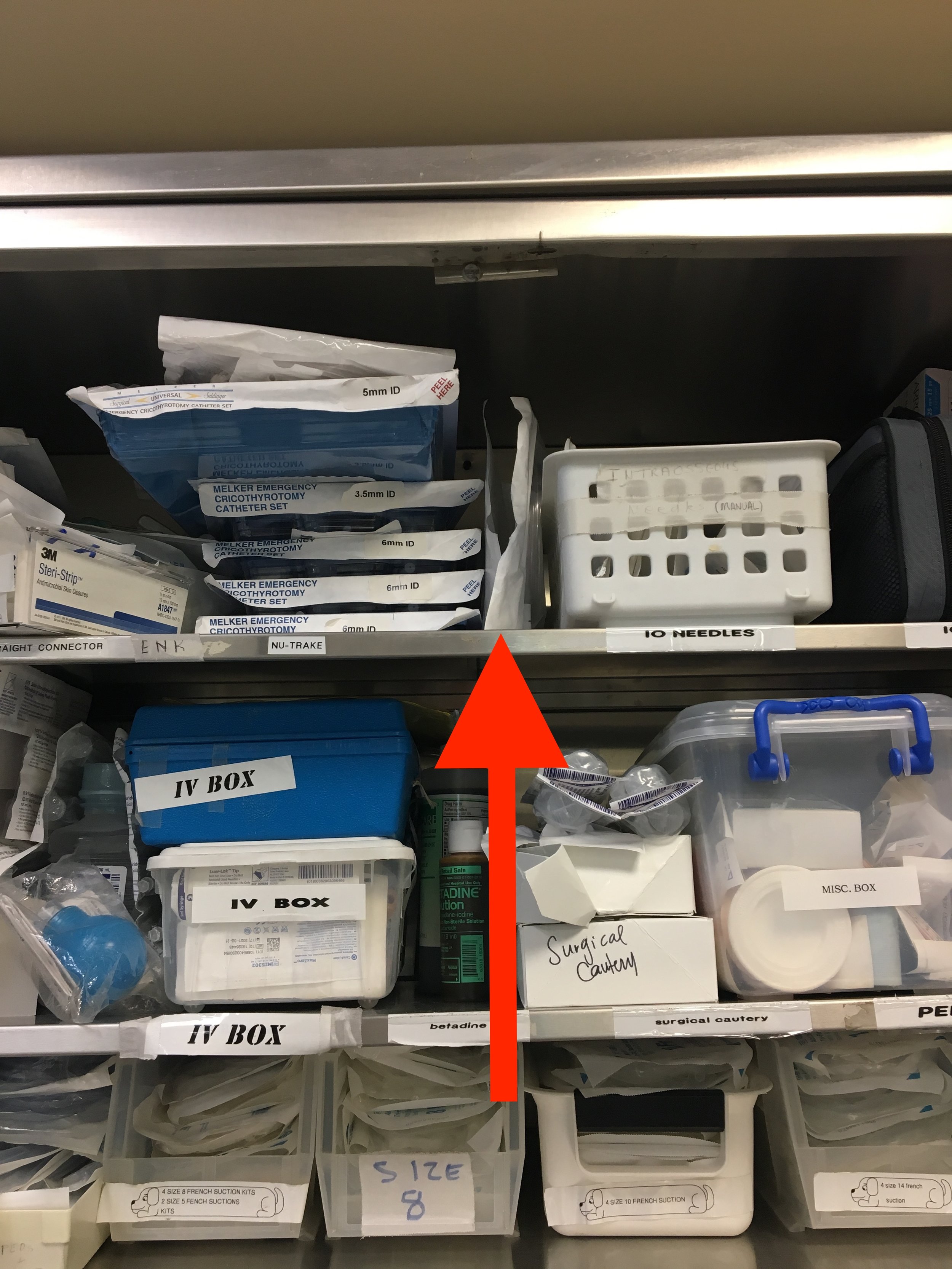
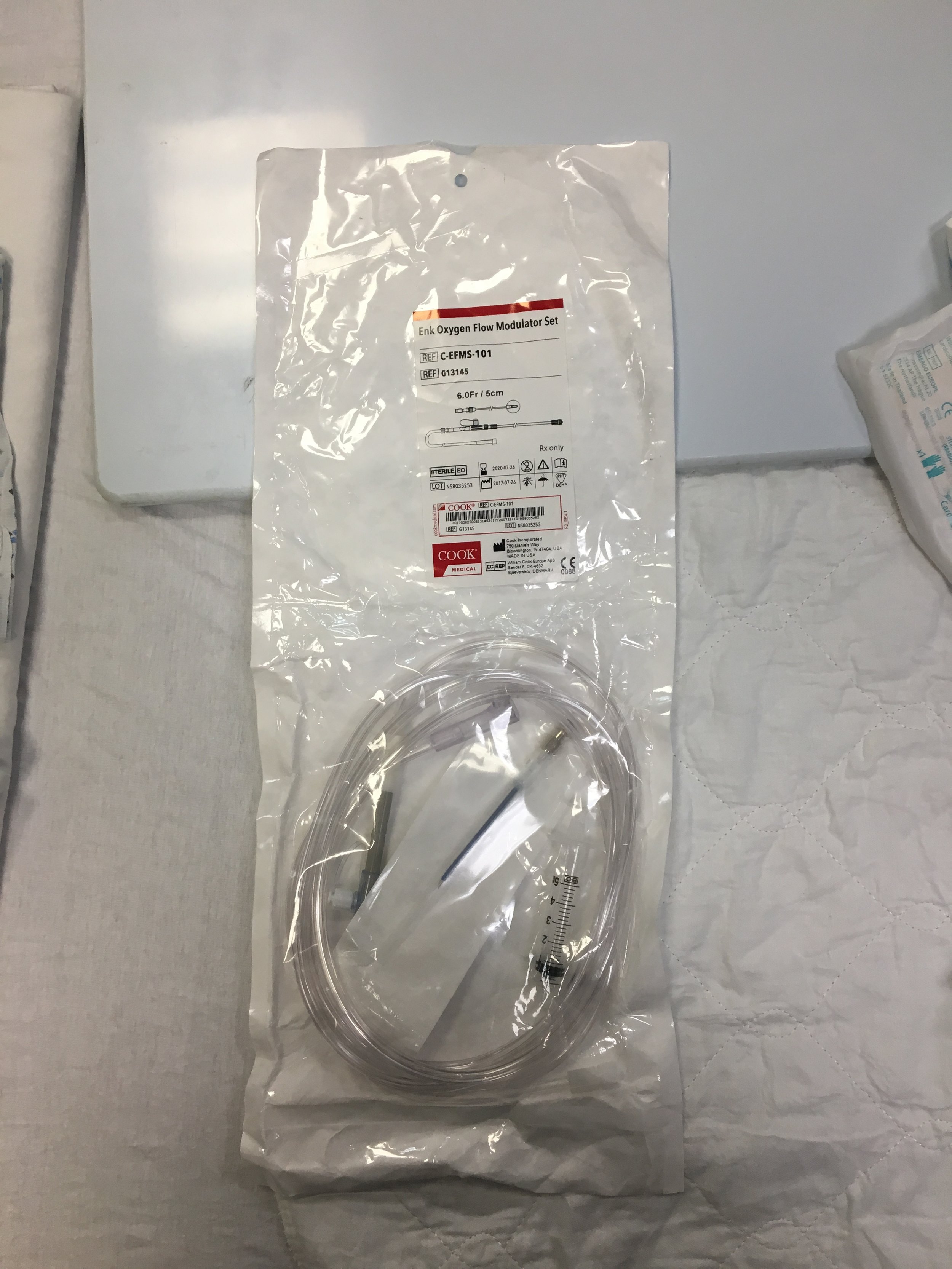
- Our kit at Maimonides is located in the Peds ED Room 30 cabinet top shelf next to the surgical cricothyroidotomy trays

IMG_7914.jpg
IMG_7908.jpg
PastedGraphic-1.tiff
PastedGraphic-3.tiff
PastedGraphic-2.tiff
IMG_7909.jpg

Steps:

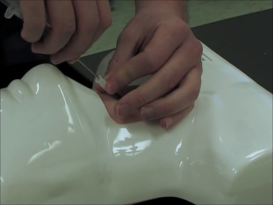
- Palpate landmarks

vlcsnap-2018-08-20-16h09m27s386.png

- Clean

- Connect tubing

vlcsnap-2018-08-20-16h08m44s472.png

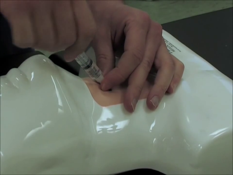
- Enter at a 45 degree angle, advance while aspirating through a 5cc syrige filled with normal saline

vlcsnap-2018-08-20-16h10m07s835.png

- Stop advancing when you see bubbles

- Advance the catheter while keeping the needle stationary

vlcsnap-2018-08-20-16h10m17s368.png

- Confirm placement in the trachea by aspirating more air from the catheter directly

vlcsnap-2018-08-20-16h10m36s892.png

- Connect tubing, O2 should be at 15 L/min

- Ventilate by covering the holes for 2-4 seconds at a time allowing for longer periods of expiration (chest recoil) to decrease risk of barotrauma (some sources recommend ratio of 1:5 covered:uncovered)

vlcsnap-2018-08-20-16h11m18s573.png

Notes:

- Transtracheal catheter ventilation BUYS YOU TIME

- Transtracheal catheter “ventilation" does NOT ventilate, it only oxygenates; CO2 will build up

- ENT should perform a surgical tracheostomy or other airway secured within 30 minutes

- It is acceptable to use the spaces in between the tracheal rings if unable to identify the cricothyroid membrane or unable to achieve enough neck extension to make accessing it feasible, as is sometimes the case with infants and young children

- A similar set-up to the above kit can be improvised as follows: BVM—>ETT adapter—>3cc syringe (plunger removed)—>14 gauge angiocath —>patient’s neck

References:

- Okada Y, Ishii W, Sato N, Kotani H, Iiduka R. Management of pediatric “cannot intubate, cannot oxygenate.” Acute Medicine & Surgery. 2017;4(4):462-466. doi:10.1002/ams2.305.

- UpToDate: Needle cricothyroidotomy with percutaneous transtracheal ventilation

https://www.youtube.com/watch?v=kDL1Y3XlFaQ

 (Video demonstration using the Cook Enk Flow Modulator kit)

 ·