EMS Protocol of the Week - A. Fib / A. Flutter / SVT

Don’t tell A FIB about SVT. You’ll make my heart FLUTTER.

We're interrupting your weekly emails from EMS extraordinaire Dr. Dave Eng to bring you some guest posts. This week we will be discussing EMS protocols for three tachyarrhythmias: 1) A.fib + A.flutter, and 2) SVT

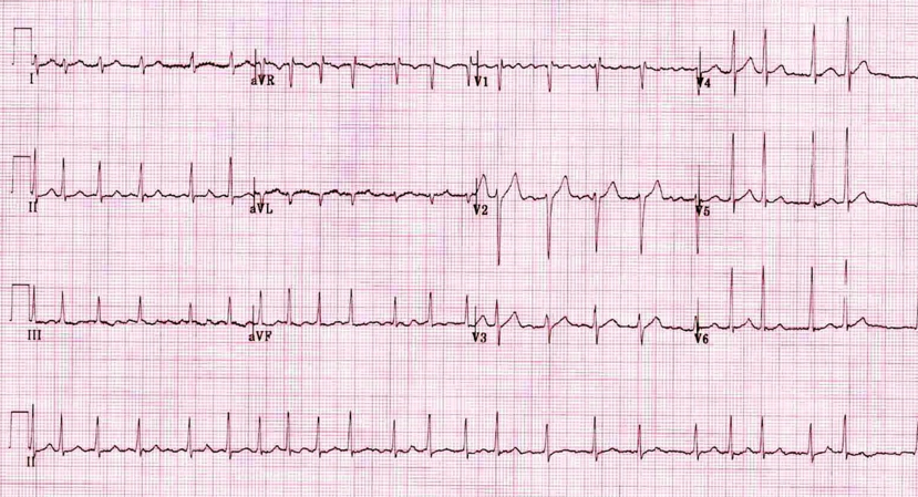
1) Atrial Fibrillation / Atrial Flutter

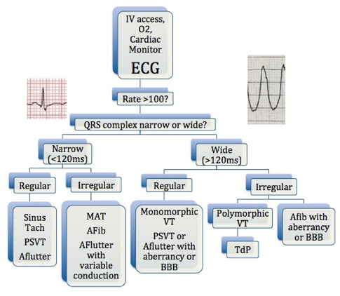
First question: is this patient stable or unstable? If this patient is hypotensive, altered, or has signs of hypoperfusion, this is an unstable patient. Standing Order will allow paramedics on scene to perform SYNCHRONIZED CARDIOVERSION up to 4 times (first 100J, then 200J, then 300J, then 360J). If that does not work, they will call OLMC for one of two options: administration of Amiodarone 150mg IV or repeating SYNCHRONIZED CARDIOVERSION at max joules setting.

If the patient is stable, there are no Standing Order available so paramedics will call OLMC for one of three options: IVF 10 ml/kg IV, Diltiazem 0.25 mg/kg IV, or Amiodarone 150mg IV. Before authorizing, first assess whether their tachyarrhythmia is compensatory for another cause (i.e. hypovolemia, sepsis, etc.) that may be better addressed first before addressing the rhythm. Choosing what to authorize is dealer’s choice, but typically IVF or Diltiazem is the safest. Diltiazem is great if there is a narrow-complex tachycardia in an otherwise stable patient. I’ve successfully converted a patient with Diltiazem who subsequently arrived at our ED in normal sinus rhythm 15 minutes later. Amiodarone is another option, however has some major side affect profiles as we know. Thoughts are it might help control rhythm while being gentler on the blood pressure in comparison to Diltiazem.

2) SVT

First question again: stable or unstable? If unstable, Standing Order allow paramedics to perform SYNCHRONIZED CARDIOVERSION up to 4 times (first 100J, then 200J, then 300J, then 360J). If the patient is stable, Standing Order allows administration of Adenosine 3 times (first 6mg, then 12mg, then 12mg). If these orders don’t work for both stable or unstable SVT, paramedics will contact OLMC for Diltiazem 0.25mg/kg IV or Amiodarone 150mg IV.

Check out www.nycremsco.org or the protocol binder on North Side for more.

Sincerely,

Joseph Liu, DO

Chief Resident, Emergency Medicine PGY-3

Maimonides Medical Center


Irregular Rhythms

EKG #1

What’s the rhythm?

afib.png

EKG #2

Dude, what’s the rhythm?

MAT.png

EKG #3

Home skillet, what’s the rhythm?

MAT.png

BONUS QUESTION: What is a Lewis Lead?

EKG #1 - Atrial Fibrillation

·      Super irregular! This is probably the most common irregularly irregular rhythm we see

·      You may have confused this with atrial flutter because of the coarse fib waves in V1

o   First, you’re not alone: 1999 study showed high frequency of incorrect diagnosis, even by cardiologists: https://www.ncbi.nlm.nih.gov/pubmed/10549907

o   It may not matter, since aflutter and afib are treated same way in the ED


But since you’re all nerds like me, here’s how to differentiate…

1.     The waves in atrial flutter are much more uniform than the ones above.

2.     Flutter waves are classically sawtooth morphology

3.     Remember atrial flutter can be regular. It is only irregular when you have variable conduction.

I.e. when you have two flutter waves and a QRS, then three flutter waves and a QRS, then four, then two again…

Screen Shot 2019-09-13 at 5.25.55 PM.png

EKG #2 - Multifocal Atrial Tachycardia (MAT)

·      Multiple foci in the atria triggering the AV node. Look at all the different P wave shapes!

·      Three separate P wave morphologies = diagnostic of MAT

·      The slow version of MAT is a wandering pacemaker (MAT without the T)

·      Classically presents in a patient with COPD or CHF

·      Note that this EKG has R axis deviation, perhaps from cor pulmonale

EKG #3 - Premature Atrial Contractions, Normal Sinus Rhythm

·      So this is yet another irregularly irregular EKG, and at first glance you may call this atrial fibrillation.

·      However, note all the well defined p waves – this is sinus tach

·      So why is it irregular?

·      Before those irregularly occurring QRS complexes, there is a p wave, and it looks exactly the same as all the other p waves (in height, width, and axis)

·      The SA node is firing early, depolarizing the ventricles, and creating an irregular rhythm

What is a Lewis Lead?

Ohmygod, great question, so glad you asked!

From Wikipedia: “A Lewis Lead is a modified ECG lead used to detect atrial [activity] when [it] is suspected… but is not definitively demonstrated on the standard 12 lead ECG.”

So you think someone has atrial flutter (sustained HR of 145 perhaps?) but you can’t see any p waves or sawtooth activity. 

Maybe you’re trying to discern SVT from fast sinus tach?

How can you solve this conundrum?

Lewis Lead!

1. You can do this on the monitor, you don’t need a 12lead2. Arrange the leads as seen here.  (The green one can go anywhere, it’s just an electrical ground)3. Check for atrial activity

1. You can do this on the monitor, you don’t need a 12lead

2. Arrange the leads as seen here. (The green one can go anywhere, it’s just an electrical ground)

3. Check for atrial activity

Before the Lewis Lead… (where the p waves at?)

After the Lewis Lead! (Retrograde p waves, mostly upright at the end of the QRS. No p waves prior to the QRS. This is SVT.)

Tachydysrhythmia Algorithm

Tachydysrhythmia Algorithm