VOTW: Uterus Didelphys

 ·   · 

Case: 29 yoF G1P0 presented to the ED with vaginal bleeding and abdominal pain. A transabdominal ultrasound was performed that did not show a definitive IUP. Beta-HCG was elevated at ~30,000. The providers then performed a TVUS that showed a gestational sac without a clear yolk sac and a concerning second structure (video 1). OBGYN was consulted to rule out ectopic pregnancy and their repeat US showed an IUP. However, they also identified 2 uteruses and a vaginal septum, leading them to believe that the patient had uterus didelphys.

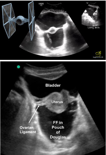
Video 1: Superficially you see a collapsed bladder. On the right side of the video, you see 1 uterine horn with a thickened endometrium and no gestational sac. On the left side of the video, towards the end of the clip, you can see an endometrium with a gestational sac. 


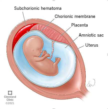
Uterus didelphys is a rare condition where a person develops 2 uteruses. It occurs when the Mullerian ducts do not fuse during embryologic development, resulting in 2 separate uteruses, each with its own fallopian tube and ovary (image 1). Some people also have 2 cervixes and 2 vaginal canals. This condition occurs in 0.3% of the population. Pregnancies in women who have uterus didelphys are considered high risk as there is less room for fetus to develop.

Case conclusion: the patient was counseled about risks and the need for close follow-up. She has a repeat ultrasound and appointment in the outpatient clinic this week.

Happy Scanning!

- Ariella Cohen, MD

References: https://my.clevelandclinic.org/health/diseases/23301-uterus-didelphys


VOTW: Subchorionic Hemorrhage

 ·   · 

Case: A 26 yoF who was 5 weeks pregnant presented to the ED after being pushed out of a parked car by her partner. She complained of wrist pain but requested an US to check on her pregnancy. She denied vaginal bleeding or pelvic pain. She had an IUP that was confirmed via US before the assault.

A transvaginal US was performed (video 1) that showed a subchorionic hemorrhage (SCH) > 50% of the gestational sac. Notebe careful not to confuse a large SCH with another gestational sac (image 1). 

Image 1

A SCH typically occurs within the first 20 weeks of gestation and is when blood accumulates between the uterine wall and the chorionic membrane (image 2). While many are found incidentally, some patient's may present with vaginal bleeding.

(What is the chorion? It is a membrane that surrounds the developing fetus along with the amnion. It eventually forms the fetal placenta and provides nourishment and protection for the developing embryo.)

On ultrasound, a SCH will typically appear as a crescentic collection with an elevation of the chorion. The echotexture can vary from hyperechoic (acute) to hypoechoic (chronic) based on the duration of the SCH. A SCH is considered large if it is > 50% of the size of the gestational sac. While many resolve during pregnancy, a large SCH can increase the risk of placental abruption, preterm labor, and miscarriage. These patients therefore require close OB follow-up for serial ultrasounds.

Case conclusion: the patient’s workup was negative, she was educated about the found SCH, and given OB follow-up later that week. 

Happy scanning!

Ariella Cohen, M.D.


References:

https://radiopaedia.org/articles/subchorionic-haemorrhage-2?lang=us

https://my.clevelandclinic.org/health/symptoms/23511-subchorionic-hematoma


VOTW: In a pelvis, far far away

27 yoF presented to the ED with acute onset abdominal pain and distention. An ultrasound was performed that showed intraperitoneal fluid. Free fluid was identified at the liver tip and in the pouch of Douglas (video 1). 

As a reminder, the pouch of Douglas is the potential space between the uterus and rectum (seen in the suprapubic view).


Video 1 shows something called the “TIE fighter sign,” where large amounts of free fluid fill the pouch of Douglas, posterior to the uterus. The uterus and ovarian ligaments are suspended between the pouch of Douglas and the bladder. This sonographic sign is based on the fact that the appearance of the uterus and ovarian ligaments looks like the “Twin Ion Engine” fighter from the Star Wars movies (Image 1).

While the free fluid was initially presumed to be from a ruptured ovarian cyst, the patient remains admitted to workup this ascites of unknown etiology

Happy scanning!

Ariella Cohen

M.D.  Emergency Medicine 

Maimonides Medical Center