VOTW: Distal Radius Fracture

This week’s VOTW is brought to you by myself!


A 72 year old female came in the ED after a FOOSH and suffered a distal radius fracture w/ dorsal angulation seen on x-ray. A POCUS was performed which showed…

Clip 1 shows the dorsal distal radius with sudden cortical disruption and dorsal angulation consistent with the fracture site. The probe marker is facing towards the hand. Clip 2 shows a hematoma block performed w/ ultrasound guidance- the needle is seen entering the fracture site precisely where the fragments meet. Reduction of the fracture was then performed once adequate analgesia was achieved.

Image 1 is prior to reduction. Image 2 is s/p first attempt at reduction. Unhappy with the alignment, reduction was attempted one more time resulting in Image 3 where the alignment is improved. Post-reduction x-rays were obtained, the patient was placed in a sugar-tong splint and discharged with orthopedic follow up.

POCUS for distal radius fractures

In a small study of 83 patients with distal radius fractures, POCUS was 98% sensitive and 98% specific for identifying the fracture when compared to x-rays. Sensitivity and specificity of POCUS ffor the need for reduction was 98% and 100% respectively (1).

While POCUS may not replace x-rays for the management of fractures, it can assist with procedural guidance for hematoma blocks and can evaluate for the adequacy of reduction in real-time rather than waiting for the x-ray tech to come around in between reduction attempts.

How to Identify a fracture

  • Use a linear high frequency probe

  • Visualize the distal radius in its long axis from multiple planes

  • Look for a disruption/angulation in the echogenic cortex

How to perform a ultrasound-guided hematoma block

  • Obtain 10ml of lidocaine drawn up in a syringe, connect it to a saline lock and an injection needle

  • Locate the fracture site using the linear probe

  • Advance the needle into the skin in-line with the probe and guide it into the fracture site

  • Have an assistant inject 10ml of lidocaine into the fracture site

References

Kozaci et al. Evaluation of the effectiveness of bedside point-of-care ultrasound in the diagnosis and management of distal radius fractures. American Journal of Emergency Medicine Volume 33, Issue 1, 2015, Pages 67-71

Happy Scanning!

Your Sono Team


VOTW: Interscalene Brachial Plexus Block

This week's VOTW is brought to you by the ultrasound team starring one of our interns!

A 16 year old male with a history of a previous shoulder dislocation presented to the Peds ED for L shoulder pain after a fall, and was found to have an anterior shoulder dislocation. The UST was paged to the bedside for a interscalene nerve block. 

In Image 1 (with probe marker directed medially) we see the anterior scalene muscle, middle scalene muscle and nestled comfortably in between in the interscalene groove is the brachial plexus which has the appearance of a "stoplight". The sternocleidomastoid muscle can also be seen superficial to the anterior scalene muscle. Image 2 shows the same images with relevant anatomy labeled

In Clip 1 we see the needle entering the neck from laterally to medially, using the in-plane approach. Spread of local anesthetic is seen within the interscalene groove. The middle scalene muscle is seen being "pushed" away from the brachial plexus

Dr. Zafrina successfully performed this nerve block and the patient underwent a shoulder reduction using external rotation. The shoulder was reduced within 3-5 seconds w/ minimal effort and the pt said "WOW, that was so much better than my last dislocation!"

Indications of the block

The interscalene nerve block provides blockade to the C5 + C6 + C7 nerve distribution (C8 and T1 are not blocked), and can provide effective analgesia for

  • proximal humerus fractures

  • shoulder dislocations

  • deltoid abscess/I&D

It does not reliably provide analgesia to more distal parts of the arm such as the elbow, nor does it block the axilla. For more distal pathology consider the supraclavicular brachial plexus block or a peripheral block (median/ulnar/radial).

Evidence?

Studies looking at the use of interscalene blocks for shoulder reductions have shown decreased length of stay compared to procedural sedation1 and the block allows us to avoid the complications associated with procedural sedation

This block also provides motor blockade and can make a shoulder reduction significantly easier by relaxing the muscles in the shoulder

Potential complications

  1. Vascular injury- The hypoechoic circular appearance of the C5-C7 nerve roots can look similar to a vessel so use color flow doppler to differentiate it from the surrounding vasculature

  2. Phrenic nerve paralysis- runs along the brachial plexus on the way to the diaphragm. Avoid the procedure in patients with severe lung disease, active respiratory discomfort or a patient with limited lung reserve. Reduce this risk by using low volume of anesthetic (10ml)

  3. As with any nerve block, calculate the max anesthetic dose for the patient, make sure your patient is always on a monitor for quick recognition of the feared complication LAST (local anesthetic systemic toxicity), and know where to find intralipid (above cabinet in resus room 53) 

How to perform the block

  1. Get a nerve block kit (cabinet in Resus 54)

  2. Get 5-10ml of local anesthetic 

    • short-acting like lidocaine for a short procedure such as I&D or shoulder reduction

    • longer-acting like ropivacaine or bupivacaine for a patient going home with a fracture

  3. place patient in lateral decubitus or place a shoulder roll to expose more of the lateral neck region

  4. place a linear probe in transverse orientation at the level of the larynx and identify the carotid and IJ (see image below)

  5. slide the probe laterally to find the sternocleidomastoid, anterior and middle scalene and the brachial plexus- look for the “stoplight”

  6. Using the in-plane approach, direct your needle to the space between the middle scalene muscle and brachial plexus, starting laterally

  7. Inject local anesthetic and watch the middle scalene be pushed off of the brachial plexus

Here is a great resource on how to perform this block from our friends at Highland: https://highlandultrasound.com/interscalene-block

References:

1. Blaivas M, Adhikari S, Lander L. A Prospective Comparison of Procedural Sedation and Ultrasound-guided Interscalene Nerve Block for Shoulder Reduction in the Emergency Department. Acad Emerg Med. 2011;18(9):922-927.

Happy Scanning and Blocking,

Your Sono Team


Unstable Pelvic Ring Fractures

The pelvic ring consists of the sacrum and two innominate bones, which are made up of the pubis, ilium, and ischium. These bones are held together by strong ligaments to give the pelvis stability.

A pelvic ring fracture is a severe fracture with 2 breaks in the circular ring, leading to an unstable pelvis and a potentially unstable patient. Fractures that disrupt the pelvic ring predispose patients to bleeding given the large network of arterial and venous anastomoses. Patients who have an isolated pelvic fracture and are hypotensive carry a mortality of 15-40%. Most vascular injuries in the pelvis are venous (90%). While rare, arterial bleeds (10%) should be suspected when a pelvic binder is placed but the patient remains hemodynamically unstable. The retroperitoneal space can accumulate 4 liters of blood before venous tamponade occurs. Pelvic binders are useful in that they can help tamponade bleeding veins, decrease total pelvic volume, and prevent the shifting of bony fragments.

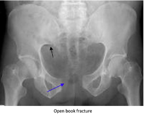
Other unstable pelvic fractures include lateral compression fractures, "open book" pelvic fractures, and vertical shear fractures. Lateral compression fractures occur when a lateral force vector (t-bone in an MVC) causes an anterior ring disruption and sacral fracture.

“Open book” fractures occur as a result of anteroposterior compression injury to the pelvis, commonly caused by high-speed trauma or elderly falls. There is a disruption to the pubic symphysis and the pelvis opens like a book. Diastasis of > 1 cm (blue arrow) can indicate instability. Disruption of the pubic symphysis, one of the strongest ligamentous structures in the human body, requires a lot of force and should be a red flag to look for other injuries to the head, spine, chest, or abdomen.

Vertical shear pelvic fractures are seen when one-half of the pelvis shifts upward as a result of a fracture of ipsilateral anterior and posterior pelvic ring fractures. They typically occur as a result of high-energy force applied in the axial direction (aka from the gas pedal to the femur and up to the pelvis). Patients may have an unstable pelvis and leg length discrepancy.

For all unstable fractures, you should appropriately resuscitate and stabilize the patient. Give blood as needed but avoid transfusing through lower limb access because it may drain into the retroperitoneal space. If there is a pelvic ring fracture, consider binding the pelvis. Your binder should lay over the greater trochanters and have enough force to close the pelvic ring (video:https://www.youtube.com/watch?v=tWLBZKeWEkg).