CXR- Consolidation or Atelectasis?

Here is a quick guide on differentiating consolidations vs atelectasis on chest x-ray.

The reason that we can differentiate structures on x-rays is due to differences in density. For example, the lungs are air-filled and appear black whereas the ribs, vertebrae, and heart are solid and appear white. 

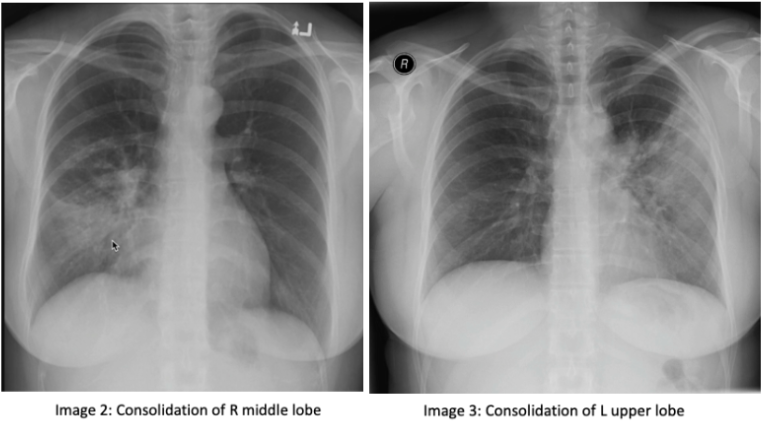
Consolidation: consolidation represents the replacement of alveolar air with fluid, blood, pus, or other substances. There are 3 lobes of the right lung, the upper, middle, and lower lobes. The right middle lobe sits next to the heart border. The left lung has 2 lobes, the upper and lower lobe. The left upper lobe sits next to the heart (image 1). If you have an obscured right heart border, it may indicate consolidation of the right middle lobe (image 2). Similarly, an obscured left heart border may indicate a consolidation in the left upper lobe (image 3). The lower lobes of each lung sit next to the hemidiaphragm. If you cannot make out a hemidiaphragm, it may suggest that there is something of similar density, such as a consolidation, in that lower lobe.

On a normal lateral chest x-ray, the vertebrae should get progressively darker as you get closer to the bases, known as the "more black sign". The vertebrae located near the apex of the lung have overlying muscles, making them appear white, compared to those at the bases that have overlying air, which makes them appear darker (image 4). You should also be able to make out 2 hemidiaphragm on the lateral x-ray with sharp costophrenic angles.

Atelectasis: Atelectasis refers to the collapse of a lung portion. On a normal x-ray, ⅓ of the heart is located on the right and ⅔ of the heart is located on the left side of the chest (image 5). In atelectasis, you will see the mediastinum shift towards the affected side due to volume loss, causing the heart and trachea to shift (image 6). In addition, the unaffected lobe on the ipsilateral side will be hyperlucent as a result of compensatory hyper-expansion. The rib spaces on the affected side may also be closer together when compared to the contralateral side and there may be an elevation of the ipsilateral hemidiaphragm. 

Tip: don’t be fooled by a rotated cxr. Rotation can be assessed by measuring the distance between the medial edges of the clavicles to the vertebral spinous processes. They should be equal or near equal.


Thanks for reading! 

Ariella 

References: 

https://radiopaedia.org/courses/emergency-radiology-course-online/pages/1417

https://radiopaedia.org/articles/lung-atelectasis


POTD: Can I Go Home With My PE?

Congrats, Maimo Fam! You ordered the correct CT and you subsequently found that Pulmonary Embolus (PE). ...Now what?

This POTD was requested for further discussion on risk stratifying patients that can potentially be discharged with a pulmonary embolus. Let's talk about the PESI Score!

Pulmonary Embolism Severity Index (PESI)

The PESI is designed to risk stratify patients who have been diagnosed with a PE in order to determine the severity of their disease. This can help physicians make decisions on the management of those patients who could potentially be treated as out-patient, as well as raise concern for those who are determined to be high-risk and could benefit from higher levels of care.

In the setting of a patient diagnosed with PE, the PESI can be utilized to determine mortality and long term morbidity. For those determined to be very low risk (score ≤ 65), all studies showed a 30-day mortality <2%. In the validation, low risk (Class I and II) had a 90-day mortality of 1.1%. The non-inferiority trial demonstrated Class I and II could have been treated as outpatients assuming no other issues.

Sounds great, but what's the catch? Although the PESI tool has been externally validated, there are a few pitfalls to be aware of.

In the setting of a patient with renal failure or severe comorbidities, clinical judgement should be used over the PESI, as these patients were excluded in the validation study.

The PESI score determines risk of mortality and severity of complications.

The score does not require laboratory variables.

It is meant to aid in decision making, not replace it. Clinical judgement should always take precedence.

The PESI score determines clinical severity and can influence treatment setting for management of PE. Class I and II patients may possibly be safely treated as outpatients in the right clinical setting.

Class I - Scores ≤ 65 indicate very low risk.

Class II - Scores of 66-85 indicate low risk.

Class III - Scores of 86-105 indicate intermediate risk.

Class IV - Scores of 106-125 indicate high risk.

Class V - Scores >125 indicate very high risk.

Again, studies show PE patients with PESI class I or II seem safe to manage as outpatients. But as always, cOrReLaTe ClInIcAlLy.

Some final thoughts:

Social situation should also be taken into account before considering outpatient management (including the appropriate administration of anticoagulants).

Given low mortality of low risk PE, outpatient management would save significant funds over hospitalization (cited as $4,500 per avoided admission).

The non-inferiority trial showed successful and safe outpatient management of Class I and II patients.

As with other tools and scores we use in the ED, use your gut and your clinical judgement. These tools are to help you in your decision, but you're the only one that can put all the pieces of your patient's clinical puzzle together. I have faith in all of you to do what's best for your patient.

References:

Aujesky D, Obrosky DS, Stone RA, et al. Derivation and validation of a prognostic model for pulmonary embolism. Am J Respir Crit Care Med. 2005;172:1041-1046.

https://www.mdcalc.com/pulmonary-embolism-severity-index-pesi

https://wikem.org/wiki/Pulmonary_embolism

 · 

POTD: Emergent Trach Complications

 Most common Tracheostomy Complaints Include the Following:

o   Dislodgement

o   Decannulation


Equipment:

o   3 parts  (past photo)

o   Outer cannula (rigid)

  • §  Top portion of the trach is called the neck plate

    ·      On the right upper hand corner you will find all the information you need in terms of sizing

  • o   Size 4, 6, 8 is the measurement of the inner diameter

o   Inner cannula

  • §  Must be inserted into the outer cannula to be able to bag the patient or connect the patient to the vent

  • §  You do not need the inner cannula if the patient is trach to air

o   Obturator  

  • §  The most distal portion of the outer cannula is blunt and has sharp edges the obturator prevents you from causing any damage when inserting the outer cannula

Important things to know when you get a tach patient

o   Size ( 4,6,8)

o   Cuffed or uncuffed

o   Reason for Trach

o   Date of placement

o   Stoma healing roughly 7-10 days

  • §  Increased risk of creating a fall passage if you replace the trach within 10days


Uncuffed trach are mostly used in patients to allow them to speak. If you need to ventilate a patient you must have a cuffed trach


Step-wise Management  of Patient with respiratory Distress in the Setting of a Trach

o   Default action for all patients in respiratory distress is to bag the face and the neck

o   High flow or PPV

o   How to bag the stoma if the trach is dislodged

o   Pediatric BVM

o   LMA (inflate a size 3 or 4  LMA and seal it around the stoma)

o   Remove the inner cannula and clean it. Replace it with either a new one or the clean one

o   Insert a sterile in-line suction catheter

o   If you can only insert the suction 1-2cm your tube is either dislodged or obstructed

o   If suctioning fails will need to deflate the cuff and push it in further and re-inflate it

o   If deflating the cuff fails will need to remove the trach tube

o   Can now intubate through the stoma or oropharynx

 Laryngectomy patient:

o   Cannot intubate through the mouth must go through the stoma

 If inserting an ET tube into the stoma only go until you loose site of the cuff then stop and inflate. Very short distance the tube needs to travel for a trach compared to an oropharyngeal intubation

 Algorithm

o   Green Algorithm (patent upper airway)

o   Red Algorithm (laryngectomy patient)

References:

o   https://www.youtube.com/watch?v=szNsOtwEU8k

o   https://emcrit.org/wp-content/uploads/2012/09/guidelines-trach-emergencies.pdf

o   https://wikem.org/wiki/Tracheostomy_complications

o   http://www.emdocs.net/trach-travails-need-to-know-ed-tricks-for-airway-emergencies-in-tracheostomy-patients/

o   https://first10em.com/tracheostomy/

 ·